

This photo gallery presents the anatomical structures found on paranasal sinuses radiography.
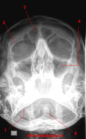
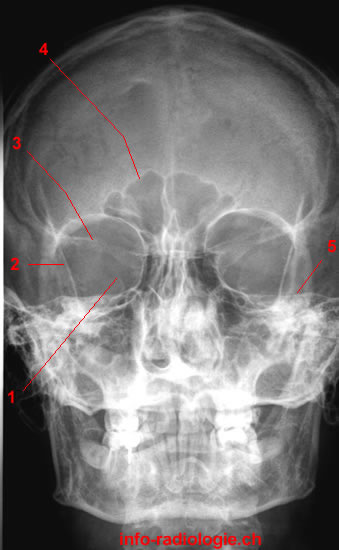
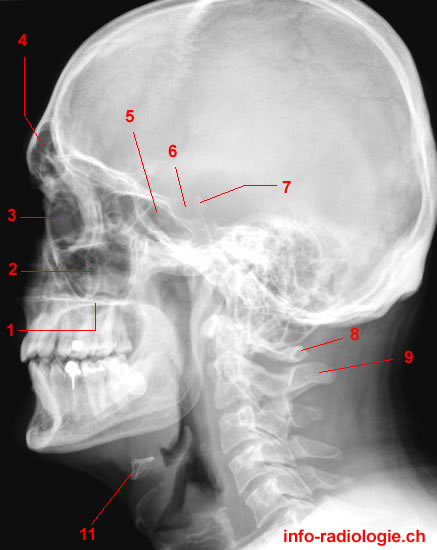
Paranasal Sinuses
Paranasal sinuses refer to paired air-filled spaces surrounding the nasal cavity(1). The nasal cavity is a system of air channels that connect the nose with the back of the throat.
Paranasal sinuses enable the circulation of the air breathed in and out of the respiratory system(2). They consist of four pairs: frontal sinuses, maxillary sinuses, ethmoidal sinuses, and sphenoidal sinuses(3). These names originate from the facial bones where the sinuses reside.
Frontal sinuses lie superior to the eyes within the frontal lobe(4). Meanwhile, maxillary sinuses—the largest of all paranasal sinuses—contain thin walls usually penetrated by the posterior maxillary teeth’s long roots. These sinuses are predominant under the eyes in the maxillary bones.
Different discrete air cells within the ethmoid bone between the nose and the eyes form ethmoidal sinuses(5). Sphenoid sinuses occupy the sphenoid bone.
Moreover, paranasal sinuses can be divided into anterior and posterior sinuses(6). The anterior sinuses include the frontal, maxillary, and anterior ethmoid air cells. Meanwhile, the posterior sinuses consist of the posterior ethmoid and sphenoid sinuses.
Although the function of paranasal sinuses remains vague, they are believed to play the following roles(7):
- Reducing the skull’s relative weight
- Enhancing the resonance of the voice
- Insulating sensitive structures from rapid temperature fluctuations in the nose
- Offering immunological defense
- Providing a buffer against facial trauma
- Humidifying and heating inspired air
Radiography of the Paranasal Sinuses
Radiography of the paranasal sinus is based on the presence of air in the sinuses(8). The radiograph can show changes in air content or alterations in translucency caused by any pathology or physiological process.
Sinus X-rays may appear normal or show infection through mucosal thickening, fluid levels, or total opacity(9).
An opaque sinus may be caused by the thickening of the bony walls, small asymmetric antra, or improper centering and rotation of the head. This type of sinus can pose difficulties in interpreting radiological appearance.
A 2002 study highlighted that paranasal sinuses radiographs are vital in identifying maxillary sinus pathology even when there is no gross pathology in the nose(10).
One can detect other pathologies, like osteomas of the sinuses, erosion of the skull base, or sinus walls by tumors, apart from sinus haziness(11). Thus, a radiograph of the paranasal sinuses is a useful modality of investigation.
However, plain film radiography of the sinuses is technically demanding, and interpretation is difficult, especially in children(12). Because of the reported cases of abnormalities in asymptomatic populations, the significance of sinus radiograph abnormalities in young patients is questionable(13).
Moreover, radiographs have limitations when showing sinonasal disease processes(14). Thus, other medical imaging tools can help assess the anatomy of paranasal sinuses.
Other Tools for Evaluating Paranasal Sinuses
Computed Tomography
Computed tomography (CT) uses various X-ray images from different angles around the body(15). CT utilizes computer processing to create cross-sectional images or slices of the bones, blood vessels, and soft tissues inside the body.
A CT scan provides a greater definition of the sinuses(16). Compared with typical radiography tools, CT is more sensitive in detecting sinus pathology, particularly within the sphenoid and ethmoid sinuses.
Moreover, a CT scan can help detect sinusitis and examine sinuses filled with thickened sinus membranes(17). CT scans can provide additional information on nasal cavity tumors, detect inflammatory disorders, and aid in surgery planning by defining anatomy.
A CT scan is ideal for examining sinus anatomy, drainage pathways, and the surrounding bony structures(18). This modality can also readily determine soft tissue and orbital extension.
Magnetic Resonance Imaging
Magnetic resonance imaging (MRI) serves as a complementary tool that helps with cases where a mass or intracranial extension is suspected(19).
MRI can help assess the extent of soft tissue or bony involvement, particularly when computed tomography is inconclusive(20).
MRI creates detailed images of the body’s organs and tissues by utilizing a magnetic field and computer-generated radio waves(21). Unlike CT, MRI does not use the ionizing radiation of X-rays(22).
An MRI scan offers the best soft-tissue contrast, which is necessary to evaluate neoplasms and complicated infections that extend beyond the sinuses(23).
Moreover, MRI can obtain multiplanar sections without disturbing the patient(24). It is also the only practical cross-sectional method for acquiring direct sagittal scans.
However, MRI has its corresponding limitations. They include high cost, long imaging times, which require sedation in most children, and the inability to directly display bony landmarks, which the endoscopic surgeon needs(25).
Thus, magnetic resonance imaging serves as a secondary or tertiary examination for some patients with sinus neoplasms or complex infectious disorders(26).