

This photo gallery presents the anatomy of the chest by means of CT (axial reconstructions – mediastinal window).


The chest or thorax is the region between the neck and diaphragm that encloses organs, such as the heart, lungs, esophagus, trachea, and thoracic diaphragm.
Computed tomography (CT) of the chest can detect pathology that may not show up on a conventional chest radiograph(1).
This medical imaging tool uses special X-ray equipment and computer technology to generate detailed pictures of the body and its structures.
CT Scan of the Chest
CT scan provides images that are more detailed than regular X-rays. This technique forms a cross-sectional image to avoid the superimposition of structures in conventional chest imaging, with an increase in attenuation sensitivity(2).
The computer can combine the images to create three-dimensional (3D) models to show the size, shape, and position of the organs inside the body.
CT scans of the chest can identify the cause of various chest symptoms, including chest pain, cough, shortness of breath, or fever.
The medical images can help determine if the patient has any of the following disorders(3):
- Infection
- Benign or malignant tumor
- Pneumonia
- Tuberculosis
- Pulmonary embolism (blocked blood flow in the lung)
- Bronchiectasis (chronic obstructive pulmonary disease)
- Interstitial and chronic lung disease
- Congenital abnormalities
- Pleural effusion (excess fluid around the lungs)
- Lung cancer
A low-dose chest CT scan produces images of sufficient quality to detect diseases and abnormalities. This type of CT scan uses a lower radiation level than a conventional chest CT scan, reducing the dose by 65% or more(4).
How Does a Chest CT Scan Work?
A chest CT scan works similarly to other X-ray exams. Various body parts absorb X-rays in different amounts, allowing doctors to distinguish body parts from one another in a CT image.
Low levels of radiation are directed through the body part examined in a regular X-ray examination. An electronic image recording plate captures soft tissues, such as the heart or lungs, and shows up in gray shades.
In a chest CT scan, several electronic X-ray detectors and X-ray beams scan more efficiently. The patient moves into a continuously rotating scanner while images are acquired per second in a spiral or helical profile(5).
The CT scanner makes many measurements through the cross-sectional plane of the thorax or chest from different rotational angles. The vast number of overlapping images improves spatial resolution in the cross-sectional image and 3D reconstructions.
Computer software processes a large volume of data to produce cross-sectional images of the body.
CT imaging is also called slices. The image slices are reassembled by the computer program, resulting in a detailed multidimensional view of the body’s interior.
Standard CT uses current technologies with the same minimum resolution as high-resolution CT. However, standard CT is reconstructed with a larger slice thickness in multiple imaging planes(6).
Thicker reconstructions make small legions, like nodules, more visible. Meanwhile, thinner slices can be reconstructed to visualize smaller structures in finer detail.
Certain CT scans require the use of contrast (iodine-based dye) into the body before the test. Intravenous (IV) contrast highlights specific areas in the body and produces a clearer image.
IV contrast may be injected into a vein in the patient’s arm or hand. However, it may not be recommended for patients with a history of anaphylactic reactions to iodine(7).
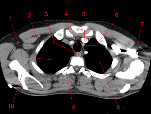
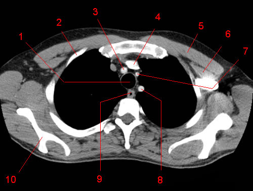
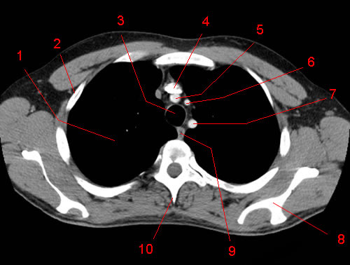
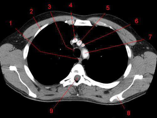
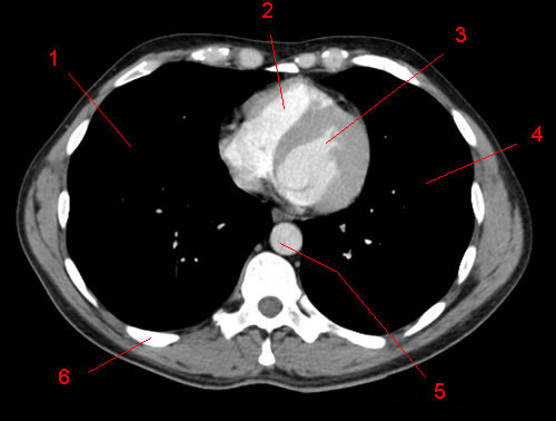
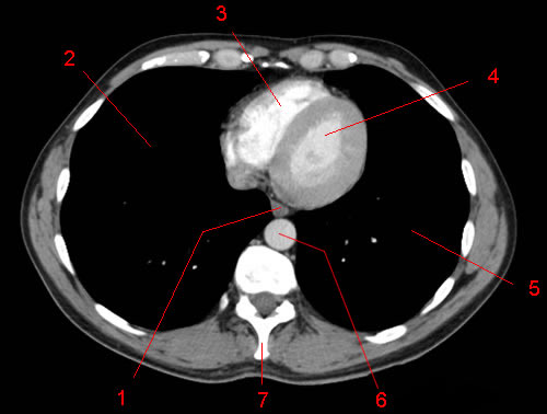
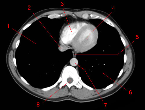
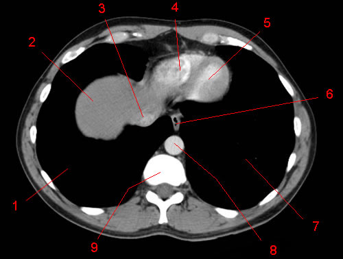
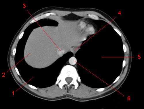
Anatomy of the Chest
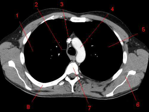
The vital structures of the thoracic cavity (chest cavity) can be identified at certain key points within the chest(8).
Great vessels region:
- Trachea
- Oesophagus
- Subclavian vessels
- Carotid vessels
- Lung apices
- Bony structures
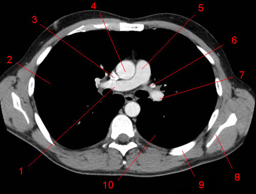
Aortic arch region:
- Superior vena cava
- Aortic arch
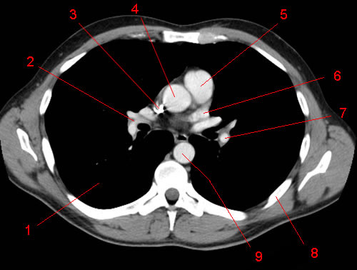
Carina and pulmonary vessel region:
- Ascending and descending aorta
- Bifurcation of the trachea
- Aortic arch
- Pulmonary arteries
- Pulmonary trunk
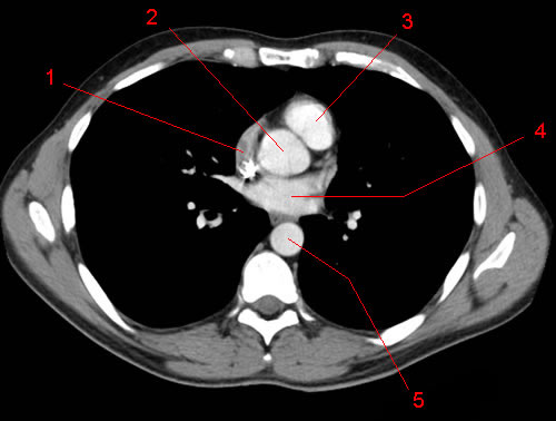
Atria region:
- Atria
- Coronary arteries
- The superficial aspects of the ventricles
Ventricular region:
- Ventricles
- Interventricular septum
- Pericardium
- Pericardial sac
- Dome of the diaphragm
Interpretation of Chest CT Scan
Below is the suggested approach for CT chest interpretation(9):
- Do a full review of the patient’s history and examination.
- Compare an individual’s previous imaging tests with their most recent scan to help with the diagnosis.
- Identify the orientation of the lung or heart images on the film. The axial image is displayed horizontally, like looking at the patient from the foot end of the bed.
- Coronal and sagittal images can be reconstructed as long as the original slices are thin and in proximity to each other.
- A systematic approach ensures the identification of abnormalities. Anatomical structures that are easily identifiable allow doctors to gain orientation.
- The imaging aids should be scrolled through to aid in anatomical differentiation and dynamic assessment.
- Whiting, P., Singatullina, N., & Rosser, J. H. (2015). Computed tomography of the chest: I. Basic principles. Bja Education, 15(6), 299-304.
- NHLBI. Chest CT Scan. Retrieved from: https://www.nhlbi.nih.gov/health-topics/chest-ct-scan
- ibid.
- Zhu, X., Yu, J., & Huang, Z. (2004). Low-dose chest CT: optimizing radiation protection for patients. American Journal of Roentgenology, 183(3), 809-816.
- Whiting, P., Singatullina, N., & Rosser, J. H. op. cit.
- ibid.
- Skinner, S. (2015). Guide to thoracic imaging. Australian family physician, 44(8), 558.
- Whiting, P., Singatullina, N., & Rosser, J. H. op. cit.
- ibid.