Partner Message: Find out more about the benefits of CBD via CBD Clinicals

Atlas of BRAIN MRI

An “overview” of the brain anatomy is offered on this page. A review of brain magnetic resonance imaging (MRI) is used as support. The anatomy of the brain is studied by means of axial, coronal and sagittal views. The MRI sequence used is a 3D gradient echo T1-weighted.

For a more detailed description of the anatomy of the brain, visit the menu entitled “MRI of the brain: region by region” which is in the right column of this page. Each region is studied with high resolution imagess and reference planes are detailed. The MRI sections come from the same patient.

Brain MRI: axial cut, T1-weighted. Image 1.
1, Medulla. 2, Cerebellar hemisphere. A, Anterior. P, Posterior.
  • Brain MRI: axial cut, T1-weighted. Image 1.
    1, Medulla. 2, Cerebellar hemisphere. A, Anterior. P, Posterior.

  • Brain MRI: axial cut, T1-weighted. Image 2.
    1, Medulla. 2, Glossopharyngeal nerve (IX). 3, Cerebellar tonsil. 4, Cerebellar hemisphere. A, Anterior. P, Posterior.

  • Brain MRI: axial cut, T1-weighted. Image 3.
    1, Temporal lobe (Right side). 2, Pons. 3, Fourth ventricle 4, Cerebellar hemisphere. A, Anterior. P, Posterior.

  • Brain MRI: axial cut, T1-weighted. Image 4.
    1, Vermis. 2, Basilar artery. 3, Globe (Right side). 4, Uncus. 5, Pons. A, Anterior. P, Posterior.

  • Brain MRI: axial cut, T1-weighted. Image 5.
    1, Cerebral peduncle. 2, Middle cerebral artery. 3, Midbrain. 4, Cerebral aqueduct. A, Anterior. P, Posterior.

  • Brain MRI: axial cut, T1-weighted. Image 6.
    1, Third ventricle. 2, Lateral sulcus. 3, Anterior commissure. 4, Pineal gland. 5, Choroid plexus (atrium of lateral ventricle). 6, Calcarine sulcus. A, Anterior. P, Posterior.

  • Brain MRI: axial cut, T1-weighted. Image 7.
    1, Insula. 2, Septum pellucidum. 3, Genu of corpus callosum. 4, Caudate nucleus. 5, Anterior arm of internal capsule. 6, Putamen. 7, Thalamus. 8, pillars of the fornix A, Anterior. P, Posterior.

  • Brain MRI: axial cut, T1-weighted. Image 8.
    1, Caudate nucleus. 2, Interhemispheric fissure. 3, Lateral ventricle. 4, Splenium of corpus callosum. A, Anterior. P, Posterior.

  • Brain MRI: axial cut, T1-weighted. Image 9.
    1, Central sulcus 2, Superior frontal gyrus. 3, Interhemispheric fissure. A, Anterior. P, Posterior.

  • Brain MRI: axial cut, T1-weighted. Image 10.
    1, Central sulcus 2, Interhemispheric fissure. 3, Cingulate sulcus (pars marginal). A, Anterior. P, Posterior.

  • Brain MRI: axial cut, T1-weighted. Image 11.
    1, Superior frontal gyrus. 2, Interhemispheric fissure. 3, Superior frontal sulcus. 4, Precentral gyrus. 5, Postcentral gyrus. 6, Central sulcus. A, Anterior. P, Posterior.

  • Image 12. Brain MRI: coronal cut, T1-weighted.1, Gyrus rectus. 2, Orbit. 3, Orbital gyrus. LF, Frontal lobe.

  • Image 13. Brain MRI: coronal cut, T1-weighted.1, Middle frontal gyrus. 2, Superior frontal gyrus. 3, Inferior frontal gyrus. 4, Temporal lobe.

  • Image 14. Brain MRI: coronal cut, T1-weighted.1, Rostrum of corpus callosum. 2, Lateral ventricle. 3, Superior frontal gyrus. 4, Superior temporal gyrus. 5, Middle temporal gyrus.

  • Image 15. Brain MRI: coronal cut, T1-weighted.1, Hippocampus. 2, Anterior commissure. 3, Column of fornix. 4, Lateral ventricle. 5, Lateral sulcus. 6, Middle temporal gyrus. 7, Inferior temporal gyrus.

  • Image 16. Brain MRI: coronal cut, T1-weighted.1, Hippocampus. 2, Caudate nucleus. 3, Corpus callosum. 4, Lateral ventricle. 5, Third ventricle. 6, Interpeduncular cistern.

  • Image 17. Brain MRI: coronal cut, T1-weighted.1, Middle cerebellar peduncle. 2, Lateral sulcus. 3, Insula. 4, Fornix. 5, Cerebral peduncle.

  • Image 18. Brain MRI: coronal cut, T1-weighted.1, Fourth ventricle 2, Superior cerebellar peduncle. 3, Lateral ventricle. 4, Fornix. 5, Colliculus. 6, Cerebellar hemisphere.

  • Image 19. Brain MRI: coronal cut, T1-weighted.1, Cerebellar hemisphere. 2, Lateral ventricle. 3, Cingulate sulcus. 4, Cingulate gyrus. 5, Choroid plexus.

  • Image 20. Brain MRI: coronal cut, T1-weighted.1, Calcarine sulcus. 2, Subparietal sulcus. 3, Cingulate sulcus. 4, Cerebellar hemisphere.

  • Image 21. Brain MRI: coronal cut, T1-weighted.1, Parietooccipital sulcus. 2, Interhemispheric fissure. 3, Cerebellar hemisphere.

  • Image 22. Brain MRI: sagittal cut, T1-weighted.1, Genu of corpus callosum. 2, Trunk of corpus callosum. 3, Cingulate sulcus (pars marginal). 4, Cingulate gyrus. 5, Splenium of corpus callosum. 6, Cerebellum. 7, Medulla. 8, Pons.

  • Image 23. Brain MRI: sagittal cut, T1-weighted.1, Caudate nucleus. 2, Lateral ventricle. 3, Cingulate sulcus. 4, Central sulcus 5, Thalamus. 6, Parietooccipital sulcus. 7, Cerebellum.

  • Image 24. Brain MRI: sagittal cut, T1-weighted.1, Caudate nucleus. 2, Lateral ventricle. 3, Parietooccipital sulcus. 4, Cerebellum.

  • Image 25. Brain MRI: sagittal cut, T1-weighted.1, Maxillary sinus. 2, Globe. 3, Caudate nucleus. 4, Cerebellum. 5, Hippocampus.

  • Image 26. Brain MRI: sagittal cut, T1-weighted.1, Gyrus parahippocampus. 2, Globe. 3, Caudate nucleus. 4, Atrium (lateral ventricle). 5, Gyrus lingual. 6, Medial occipitotemporal gyrus.

  • Image 27. Brain MRI: sagittal cut, T1-weighted.1, Hippocampus. 2, Central sulcus 3, Atrium (lateral ventricle). 4, Occipital pole. 5, Medial occipitotemporal gyrus.

  • Image 28. Brain MRI: sagittal cut, T1-weighted.1, Hippocampus. 2, Maxillary sinus. 3, Central sulcus 4, Cerebellum.

  • Image 29. Brain MRI: sagittal cut, T1-weighted.1, Temporal pole. 2, Insular cortex. 3, Central sulcus 4, Cerebellum.

  • Image 30. Brain MRI: sagittal cut, T1-weighted.1, Inferior temporal gyrus. 2, Middle temporal gyrus. 3, Superior temporal gyrus. 4, Central sulcus 5, Cerebellum.

  • Image 31 of 31. Brain MRI: sagittal cut, T1-weighted.1, Inferior temporal gyrus. 2, Middle temporal gyrus. 3, Superior temporal gyrus. 4, Lateral sulcus. 5, Central sulcus 6, Cerebellum.

Brain magnetic resonance imaging (MRI) is a common medical imaging method that allows clinicians to examine the brain’s anatomy(1). It uses a magnetic field and radio waves to produce detailed images of the brain and the brainstem to detect various conditions(2). These include tumors, inflammatory ailments, and developmental and structural abnormalities.

Given the different pathological conditions that MRI can help detect, it is vital to understand the brain’s anatomy as seen on MRI.

How Does Brain MRI Work?

Unlike X-rays or computed tomography (CT) scans, MRI does not use radiation. However, the procedure may last for 30 minutes to 2 hours(3).

Moreover, magnetic resonance imaging is often the most sensitive imaging technique in assessing the structure of the brain and spinal cord(4). MRI excites the tissue hydrogen protons, emitting electromagnetic signals back to the MRI machine. This MRI machine detects the signals’ intensity and translates the result into a gray-scale image.

Contrast agents, like gadolinium, may be given through an IV to increase the visibility of the body’s internal structures.

The most common MRI sequences used include T1-weighted (T1w) and T2-weighted (T2w) scans(5). T1w sequences display those structures mainly made with fat. Thus, they reveal gray matter as gray, white matter as white, bones as black, and cerebrospinal fluid as black. 

Meanwhile, T2w sequences highlight structures containing more water. These sequences display the gray matter as gray, white matter as darker gray, bones as black, and cerebrospinal fluid as white. 

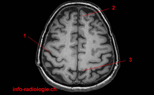
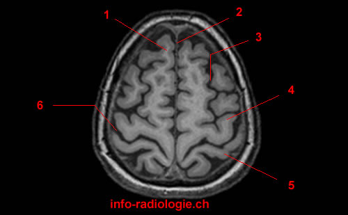
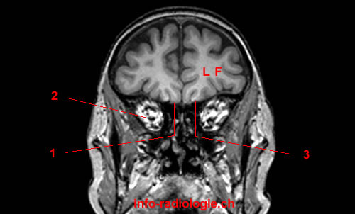
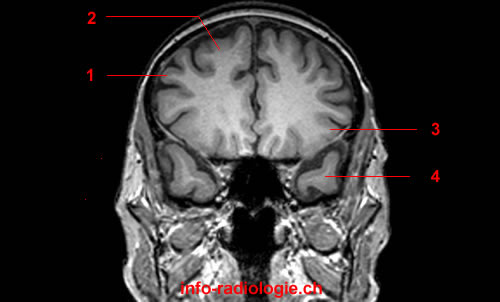
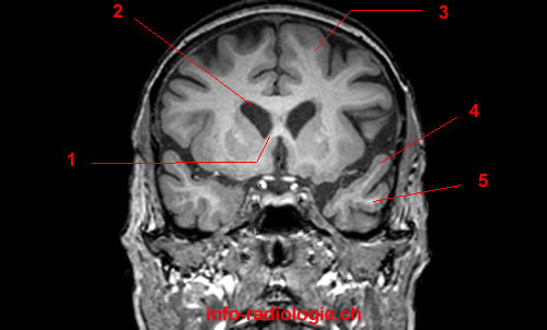
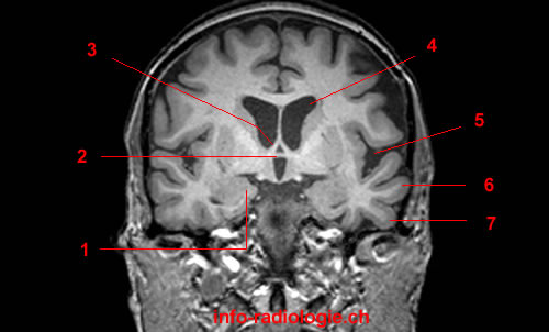
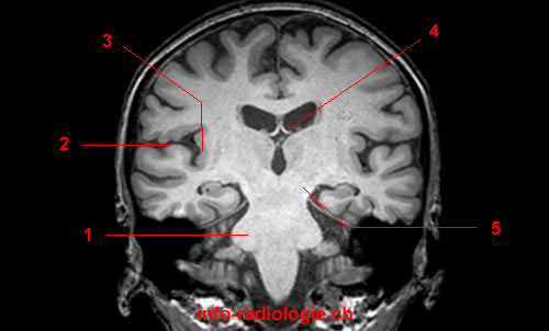
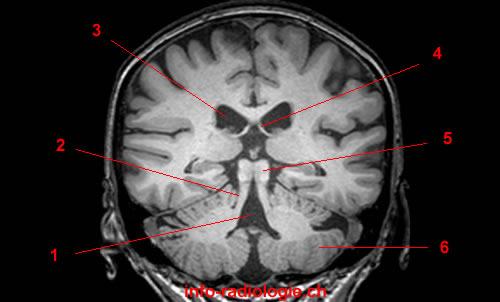
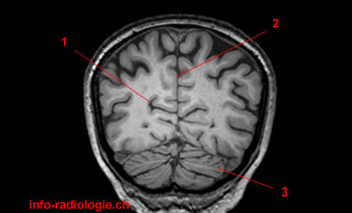
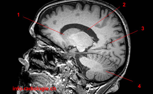
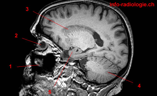
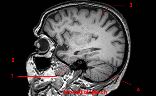
The orientation is another vital element of the scan. Three MRI orientations include axial (from top to bottom), coronal (from front to back), and sagittal (side to side) views(6).
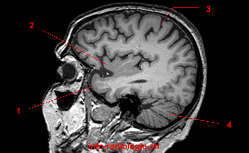
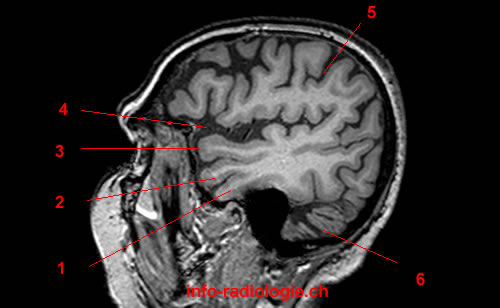
Brain Anatomy

Part of the central nervous system, the brain is an essential organ that regulates memories, thoughts, emotions, motor skills, and all other processes involved in the body(7).
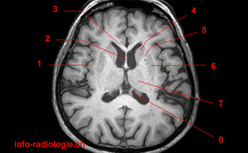
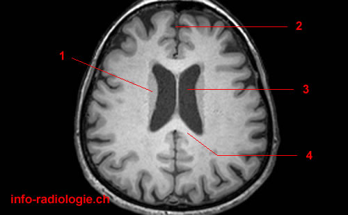
Cerebrum

The cerebrum or front of the brain consists of the right and left hemispheres(8). Its main functions include the initiation and coordination of movement, reasoning, problem-solving, emotions, learning, touch, vision, and hearing.
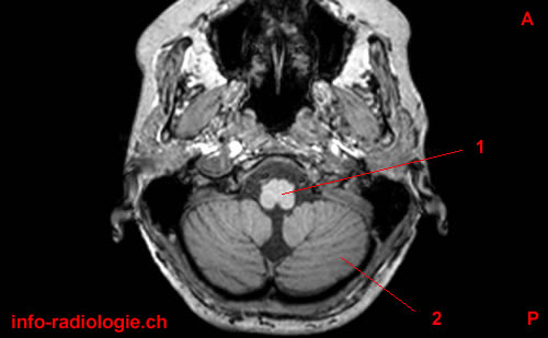
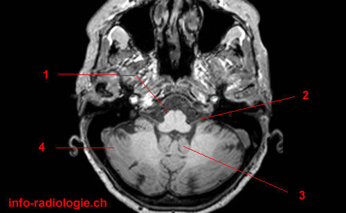
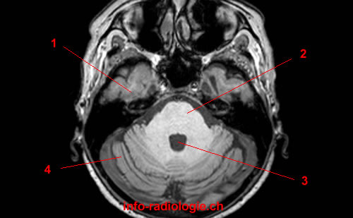
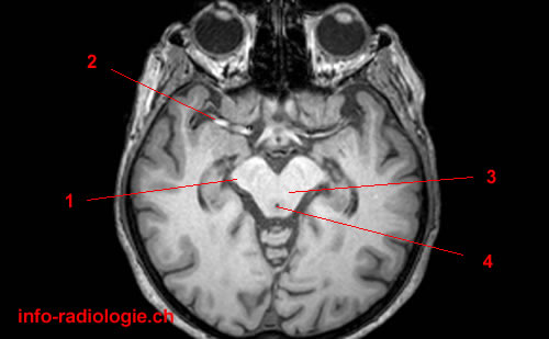
Brainstem

The brainstem is the middle part of the brain, including the medulla, pons, and midbrain(9). Its main functions include relaying sensory information, like pain, eye and mouth movement, involuntary muscle movements, respirations, hunger, consciousness, and cardiac function.

Medulla

The medulla oblongata is the lowest part of the brainstem and is the most crucial part of the brain(10). It contains important control centers for the heart and the lungs. 

Pons

Pons is a deep part of the brain located in the brainstem(11). It possesses many of the control areas for the movement of the eyes and face. 
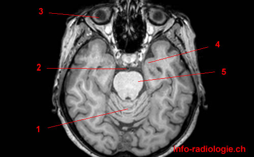
Midbrain

Midbrain refers to the shortest segment of the brainstem(12). However, it has several structures necessary to maintain different body functions, such as processing auditory and visual information and controlling movements of the eyes and face.

Cerebellum

The cerebellum is the back of the brain located at the back of the head(13). Its main functions include coordinating voluntary muscle movements and maintaining posture and balance.

Brain Lobes

Some pathological conditions may be observed from specific brain lobes(14). These brain lobes consist of the frontal lobe, parietal lobe, occipital lobe, temporal lobe, limbic lobe, and insular lobe.
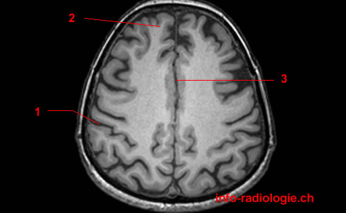
Frontal Lobe

The frontal lobe refers to the brain’s largest section located at the front of the head(15). Moreover, the frontal lobe is responsible for personality characteristics, movement, and recognition of smell.

Parietal Lobe

The parietal lobe is the middle part of the brain that helps individuals recognize objects and comprehend spatial relations(16). This lobe is responsible for pain interpretation and touch.

Occipital Lobe

The back part of the brain — the occipital lobe — is involved with vision(17)

Temporal Lobe

The temporal lobe refers to the brain’s sides responsible for short-term memory, musical rhythm, and speech(18).

Limbic Lobe

Lying deep in the parietal and frontal lobes, the limbic lobe is a functional unit usually called the limbic system(19). It helps influence bodily functions, like memory, learning, and behavior(20).

Insular Lobe

Lying lateral to the extreme capsule of basal ganglia, the insular lobe is a small part of the cerebral cortex found deep within the meeting point of the temporal, parietal, and frontal lobes(21). The insular lobe plays a role in autonomic control,  pain processing, and taste perception(22).

Different Conditions That Brain MRI Can Detect 

 Brain MRI may help detect specific abnormalities or conditions in the brain(23). These include: 

  • Tumors
  • Aneurysms
  • Congenital abnormalities
  • Hemorrhage, or bleeding into the brain or spinal cord
  • Hydrocephalus or fluid in the brain
  • Degenerative diseases, multiple sclerosis, encephalomyelitis (inflammation of the brain or spinal cord), and hypoxic encephalopathy (dysfunction of the brain caused by lack of oxygen)

Thus, understanding the brain anatomy and how MRI works can help one learn MRI’s essential role in detecting brain-related conditions.   


  1. Vasković, J. (2020, Oct. 29). Normal brain MRI. Retrieved from https://www.kenhub.com/en/library/anatomy/normal-brain-mri 
  2. KidsHealth from Nemours. Magnetic Resonance Imaging (MRI): Brain. Retrieved from https://kidshealth.org/en/parents/mri-brain.html 
  3. Johns Hopkins Medicine. Magnetic Resonance Imaging (MRI) of the Spine and Brain. Retrieved from https://www.hopkinsmedicine.org/health/treatment-tests-and-therapies/magnetic-resonance-imaging-mri-of-the-spine-and-brain 
  4. Vasković, J. (2020). Op Cit. 
  5. Ibid.
  6. Padmanaban, Sriramakrishnan & Thiruvenkadam, Kalaiselvi & T., Padmapriya & Thirumalaiselvi, M. & KUMAR, RAM. (2020). A Role of Medical Imaging Techniques in Human Brain Tumor Treatment. 8. 565-568. 10.35940/ijrte.D1105.1284S219. https://www.researchgate.net/figure/MRI-planes-for-MRI-head-scan-a-Axial-b-Coronal-c-Sagittal-MR-scanner-can-generate_fig2_338448026#:~:text=The%20basic%20planes%20of%20MRI,the%20head%20from%20the%20feet.&text=A%20sagittal%20is%20a%20Y%2DZ%20plane%2C%20which%20separates%20left%20from%20right 
  7. Johns Hopkins Medicine. Op Cit. 
  8. Ibid. 
  9. Ibid. 
  10. Ibid. 
  11. Ibid. 
  12. Vasković, J. (2020, Oct. 29). Midbrain (Mesencephalon). Retrieved from https://www.kenhub.com/en/library/anatomy/midbrain-pons-gross-anatomy 
  13. Johns Hopkins Medicine. Op Cit. 
  14. Ibid. 
  15. Ibid. 
  16. Ibid.
  17. Ibid. 
  18. Ibid. 
  19. Vasković, J. (2020, Oct. 29). Normal brain MRI. Op Cit. 
  20. Haines, D., & Mihailoff, G.A. (2018). Chapter 16 – The Telencephalon. Haines, D. E., & Mihailoff, G. A., Eds.), Fundamental neuroscience for basic and clinical applications. (pp. 225–240). Elsevier. doi.org/10.1016/B978-0-323-39632-5.00016-5. https://www.sciencedirect.com/science/article/pii/B9780323396325000165   
  21. Vasković, J. (2020, Oct. 29). Normal brain MRI. Op Cit.
  22. McGinn M.J. (2011) Insular Lobe. In: Kreutzer J.S., DeLuca J., Caplan B. (eds) Encyclopedia of Clinical Neuropsychology. Springer, New York, NY. doi.org/10.1007/978-0-387-79948-3_325. https://link.springer.com/referenceworkentry/10.1007%2F978-0-387-79948-3_325 
  23. Johns Hopkins Medicine. Op Cit. 

Need more?