Computed tomography (CT) scans of the wrist have been used to assess fusion procedures, foreign bodies, masses, and carpal fractures(1). A study suggested that CT scans allow the imaging of the wrist bones during motion(2). This procedure is essential in the diagnosis of vital carpal instabilities(3).
Wrist Anatomy
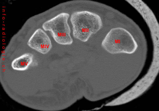
The wrist or carpus is the joint between the metacarpal joints of the hands(4). The metacarpal is any of the tubular bones between the wrist bones and each forelimb digit in vertebrates.
The forelimb has three digits (II, III and IV), each with a complete metacarpal, proximal, middle and distal phalanx.
The metacarpals form a transverse arch, allowing the thumb and fingertips to function together for better manipulation.
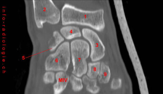
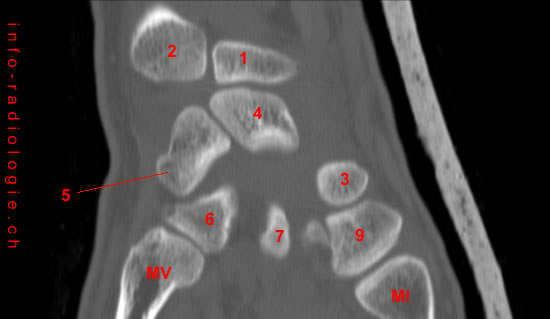
The carpus (wrist) is a complex group of bones collectively known as carpal bones(5). It is where the ulna (the small bone in the pinky side of the arm) and the radius (the large bone in the thumb side of the arm) convene(6).
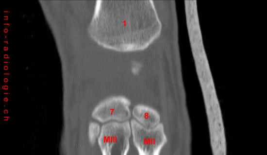
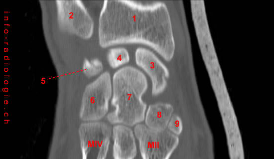
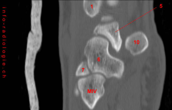
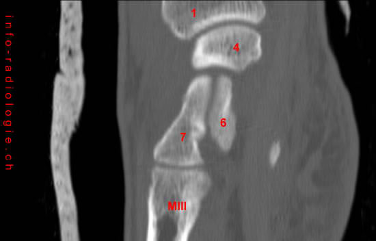
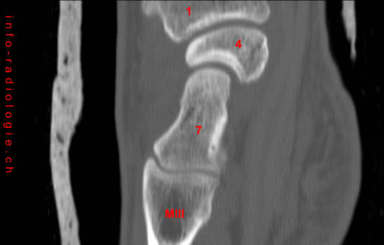
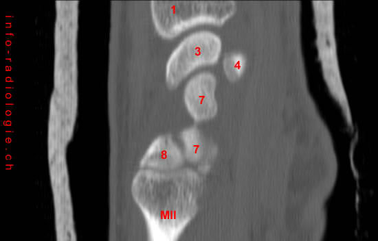
There are eight bones included in the carpal bones that are divided into proximal (nearest to the center) and distal (farthest from the center) rows(7).
Carpal bones at the upper end of the wrist(8):
- Pisiform
- Lunate
- Scaphoid
- Triquetrum
Carpal bones at the lower side of the hand(9):
- Capitate
- Hamate
- Trapezium
- Trapezoid
The scaphoid bone is the largest carpal bone and crosses the proximal and distal rows. Both scaphoid and lunate bones connect the radius and ulna bones, resulting in wrist joint formation.
Significance of CT Scan in Wrist Injuries
CT scans help detect internal injuries, diagnose bone and muscle disorders, and detect the tumor’s location, blood clots, or internal bleeding(10).
Moreover, this radiological procedure can assess whether or not a cancer treatment is effective, serving as a guide for biopsies, radiation therapies, and surgeries(11).
Aside from bone fractures, a computed tomography (CT) scan can show injuries in the soft tissues and blood vessels(12). It can also expose some injuries that an X-ray may have missed(13).
A study cited that a single shot CT improves the detection of fractures in patients with suspected wrist injuries(14).
The same study mentioned that CT scans are more accessible in modality and provide high contrast image information of bone surfaces(15).
There are various views and positions in examining the wrist using a CT scan. These views include(16):
- Wrist in the prone position
- perpendicular to the CT gantry
- PA (posterior-anterior) wrist image reformatted CT along the axis of the scaphoid bone
- Wrist in the prone position with an internal and external deviation of the hand
The majority of wrist fractures involve the ulna and the radius(17). However, the wrist fractures associated with the scaphoid bone (carpal bone at the base of the thumb) are more alarming(18).
According to the study published in the Polish Journal of Radiology, the PA position during the wrist CT scan is the view recommended in the interpretation of the distal radioulnar and intercarpal articulations(19).
This position provides a simplified image interpretation of the hamate hook that is not seen using conventional radiographs(20).
A CT scan image that uses an intravenous (IV) contrast solution can show the fluid collections and infections, as well as the vascular structure and integrity(21).
Trauma, bone lesions, humpback deformity of the scaphoid, carpal dislocation, arthritis, and scaphoid fractures are some of the wrist disorders and injuries that may require radiological imaging considerations(22).
Wrist Injuries
Individuals doing sports, performing repetitive wrist motions, and those with rheumatoid arthritis face a high risk of experiencing wrist pain(23).
The severity of wrist pain may vary depending on its cause. Injuries may be due to repetitive stress, sudden impacts, or a result of long-term diseases, like arthritis (swelling or tenderness of the joints)(24).
Wrist Fractures
Wrist fractures happen when any of the bones in the wrist break(25). The most common wrist fracture occurs in the radius bone(26).
Some wrist fractures can be stable. Non-displaced breaks, in which bones do not move out of place initially, can be stable. Displaced breaks, in which bones need to be put back to its right position, can also be stable for a cast or splint(27).
Unstable wrist fractures can cause the bones to shift into an inferior position and may need a cast to heal. This kind of fracture may tend to make the wrist look crooked(28).
A broken wrist may induce swelling, bruising, and tenderness(29). Stiffness, nerve damage, and osteoarthritis are complications that may lead to a wrist fracture(30).
Carpal Tunnel Syndrome
The pinching of the median nerve within the wrist is called carpal tunnel syndrome(31). This disorder results in the tingling and numbness of the hand, wrist pain, weak grip, and incoordination(32).
A study used a CT scan to evaluate two corpses, two healthy people, and 20 patients with carpal tunnel syndrome.
CT scans of these subjects revealed the changes in the carpal canal (a narrow passageway in the anterior of the wrist) that leads to median nerve compression, causing carpal tunnel syndrome(33).
The changes that were shown by the CT scan images included the increase in the volume of the tissues within the carpal canal, recurrent fibrosis (pathological wound healing), and the thickening of the transverse carpal ligament(34).
Conclusion
The carpus or the wrist is a complex bone structure that connects the forearm and the hand(35). It may be assessed through radiological imaging tests, such as a CT scan.
Wrist injuries may be diagnosed through imaging tests, X-rays, physical examinations, and blood tests(36). The treatments of these injuries may involve wearing a cast or wrist brace, resting the wrist, physical therapy, or surgery(37).