Partner Message: Find out more about the benefits of CBD via CBD Clinicals

Shoulder MR Arthrography Anatomy

This webpage presents the anatomical structures found on MR arthrography of the shoulder.

MR arthrography of the shoulder: coronal view, T1 FATSAT. Image 1. 1, Deltoid muscle. 2, Coracoid process. 3, Clavicle. 4, Trapezius muscle.

  • MR arthrography of the shoulder: coronal view, T1 FATSAT. Image 1. 1, Deltoid muscle. 2, Coracoid process. 3, Clavicle. 4, Trapezius muscle.

  • MR arthrography of the shoulder: coronal view, T1 FATSAT. Image 2. 1, Deltoid muscle. 2, Coracoid process. 3, Clavicle. 4, Trapezius muscle.

  • MR arthrography of the shoulder: coronal view, T1 FATSAT. Image 3. 1, Deltoid muscle. 2, Subscapularis muscle. 3, Coracoid process. 4, Clavicle. 5, Trapezius muscle.

  • MR arthrography of the shoulder: coronal view, T1 FATSAT. Image 4. 1, Deltoid muscle. 2, Humeral head. 3, Subscapularis muscle. 4, Coracoid process. 5, Supraspinatus muscle. 6, Clavicle. 7, Trapezius muscle.

  • MR arthrography of the shoulder: coronal view, T1 FATSAT. Image 5. 1, Deltoid muscle. 2, Biceps tendon, long head. 3, Humeral head. 4, Subscapularis muscle. 5, Labrum. 6, Coracoid process. 7, Supraspinatus muscle. 8, Acromioclavicular joint. 9, Trapezius muscle.

  • MR arthrography of the shoulder: coronal view, T1 FATSAT. Image 6. 1, Deltoid muscle. 2, Biceps tendon, long head. 3, Humeral head. 4, Glenoid. 5, Subscapularis muscle. 6, Supraspinatus muscle. 7, Acromion. 8, Clavicle. 9, Trapezius muscle.

  • MR arthrography of the shoulder: coronal view, T1 FATSAT. Image 7. 1, Deltoid muscle. 2, Humeral head. 3, Glenoid. 4, Supraspinatus muscle. 5, Acromion. 6, Trapezius muscle.

  • MR arthrography of the shoulder: coronal view, T1 FATSAT. Image 8. 1, Deltoid muscle. 2, Humeral head. 3, Glenoid. 4, Supraspinatus muscle. 5, Acromion. 6, Trapezius muscle.

  • MR arthrography of the shoulder: coronal view, T1 FATSAT. Image 9. 1, Deltoid muscle. 2, Humeral head. 3, Glenoid. 4, Supraspinatus muscle. 5, Acromion. 6, Trapezius muscle.

  • MR arthrography of the shoulder: coronal view, T1 FATSAT. Image 10. 1, Deltoid muscle. 2, Humeral head. 3, Glenoid. 4, Supraspinatus muscle. 5, Acromion. 6, Trapezius muscle.

  • MR arthrography of the shoulder: coronal view, T1 FATSAT. Image 11. 1, Deltoid muscle. 2, Humerus. 3, Teres major muscle. 4, Teres minor muscle. 5, Glenoid. 6, Humeral head. 7, Infraspinatus muscle. 8, Acromion. 9, Trapezius muscle.

  • MR arthrography of the shoulder: coronal view, T1 FATSAT. Image 12. 1, Deltoid muscle. 2, Humerus. 3, Triceps muscle, long head. 4, Teres major muscle. 5, Greater tuberosity. 6, Scapula. 7, Infraspinatus muscle. 8, Acromion.

  • MR arthrography of the shoulder: coronal view, T1 FATSAT. Image 13. 1, Deltoid muscle. 2, Humerus. 3, Triceps muscle, long head. 4, Teres major muscle. 5, Scapula. 6, Humeral head. 7, Acromion. 8, Infraspinatus muscle.

  • MR arthrography of the shoulder: coronal view, T1 FATSAT. Image 14. 1, Deltoid muscle (lateral). 2, Triceps muscle, long head. 3, Teres major muscle. 4, Scapula. 5, Infraspinatus muscle. 6, Acromion.

  • MR arthrography of the shoulder: coronal view, T1 FATSAT. Image 15. 1, Triceps muscle, long head. 2, Triceps brachial muscle, lateral head. 3, Deltoid muscle (lateral). 4, Teres major muscle. 5, Teres minor muscle. 6, Infraspinatus muscle. 7, Deltoid muscle (posterior).

  • MR arthrography of the shoulder: coronal view, T1 FATSAT. Image 16. 1, Triceps brachial muscle. 2, Teres major muscle. 3, Deltoid muscle. 4, Teres minor muscle. 5, Infraspinatus muscle.

  • MR arthrography of the shoulder: axial view, T1 FATSAT. Image 1. 1, Clavicle. 2, Supraspinatus muscle. 3, Scapular spine 4, Deltoid muscle.

  • MR arthrography of the shoulder: axial view, T1 FATSAT. Image 2. 1, Clavicle. 2, Supraspinatus muscle. 3, Scapular spine 4, Deltoid muscle.

  • MR arthrography of the shoulder: axial view, T1 FATSAT. Image 3. 1, Clavicle. 2, Supraspinatus muscle. 3, Acromion. 4, Deltoid muscle.

  • MR arthrography of the shoulder: axial view, T1 FATSAT. Image 4. 1, Clavicle. 2, Supraspinatus muscle. 3, Scapular spine. 4, Deltoid muscle. 5, Acromion.

  • MR arthrography of the shoulder: axial view, T1 FATSAT. Image 5. 1, Clavicle. 2, Supraspinatus muscle. 3, Scapular spine. 4, Infraspinatus muscle. 5, Deltoid muscle.

  • MR arthrography of the shoulder: axial view, T1 FATSAT. Image 6. 1, Clavicle. 2, Supraspinatus muscle. 3, Scapular spine. 4, Infraspinatus muscle. 5, Deltoid muscle. 6, Humeral head.

  • MR arthrography of the shoulder: axial view, T1 FATSAT. Image 7. 1, Deltoid muscle (anterior). 2, Glenoid. 3, Supraspinatus muscle. 4, Infraspinatus muscle. 5, Deltoid muscle (posterior). 6, Humeral head.

  • MR arthrography of the shoulder: axial view, T1 FATSAT. Image 8. 1, Coracoid process. 2, Superior glenohumeral ligament. 3, Humeral head. 4, Superior labrum. 5, Deltoid muscle (posterior). 6, Infraspinatus muscle. 7, Glenoid.

  • MR arthrography of the shoulder: axial view, T1 FATSAT. Image 9. 1, Coracoid process. 2, Labrum, anterosuperior. 3, Humeral head. 4, Deltoid muscle (posterior). 5, Infraspinatus muscle.

  • MR arthrography of the shoulder: axial view, T1 FATSAT. Image 10. 1, Subscapularis muscle. 2, Glenoid. 3, Coracoid process. 4, Deltoid muscle (anterior). 5, Humeral head. 6, Labrum, posterosuperior. 7, Deltoid muscle (posterior). 8, Infraspinatus muscle.

  • MR arthrography of the shoulder: axial view, T1 FATSAT. Image 11. 1, Subscapularis muscle. 2, Glenoid. 3, Coracoid process. 4, Middle glenohumeral ligament. 5, Humeral head. 6, Deltoid muscle (posterior). 7, Infraspinatus muscle.

  • MR arthrography of the shoulder: axial view, T1 FATSAT. Image 12. 1, Subscapularis muscle. 2, Glenoid. 3, Coracoid process. 4, Deltoid muscle (anterior). 5, Humeral head. 6, Deltoid muscle (posterior). 7, Infraspinatus muscle.

  • MR arthrography of the shoulder: axial view, T1 FATSAT. Image 13. 1, Subscapularis muscle. 2, Labrum, anterior. 3, Transverse ligament. 4, Deltoid muscle. 5, Labrum, posterior. 6, Teres minor muscle. 7, Infraspinatus muscle. 8, Glenoid. Arrow, Articular cartilage.

  • <p><strong>MR arthrography of the shoulder: axial view, T1 FATSAT. Image 13. </strong> <span class="renfort">1,</span> Subscapularis muscle. <span class="renfort">2,</span> Labrum, anterior. <span class="renfort">3,</span> Transverse ligament. <span class="renfort">4,</span> Deltoid muscle. <span class="renfort">5,</span> Labrum, posterior. <span class="renfort">6,</span> Teres minor muscle. <span class="renfort">7,</span> Infraspinatus muscle. <span class="renfort">8,</span> Glenoid. <span class="renfort">Arrow,</span> Articular cartilage.</p>

  • MR arthrography of the shoulder: axial view, T1 FATSAT. Image 15. 1, Biceps tendon, long head. 2, Humerus. 3, Deltoid muscle. 4, Triceps brachial muscle (long head). 5, Teres major muscle. 6, Teres minor muscle. 7, Infraspinatus muscle. 8, Subscapularis muscle. 9, Inferior glenohumeral ligament.

  • MR arthrography of the shoulder: axial view, T1 FATSAT. Image 16. 1, Biceps tendon, long head. 2, Humerus. 3, Deltoid muscle. 4, Triceps brachial muscle (long head). 5, Teres major muscle. 6, Teres minor muscle. 7, Infraspinatus muscle. 8, Subscapularis muscle.

  • MR arthrography of the shoulder: axial view, T1 FATSAT. Image 17. 1, Biceps tendon, long head. 2, Humerus. 3, Deltoid muscle. 4, Triceps brachial muscle (long head). 5, Teres major muscle. 6, Teres minor muscle. 7, Infraspinatus muscle. 8, Scapula. 9, Subscapularis muscle.

  • MR arthrography of the shoulder: axial view, T1 FATSAT. Image 18. 1, Biceps tendon, long head. 2, Humerus. 3, Deltoid muscle. 4, Triceps brachial muscle (lateral head). 5, Triceps brachial muscle (long head). 6, Teres major muscle. 7, Teres minor muscle. 8, Scapula. 9, Infraspinatus muscle. 10, Subscapularis muscle.

  • MR arthrography of the shoulder: axial view, T1 FATSAT. Image 19. 1, Biceps tendon, long head. 2, Humerus. 3, Triceps brachial muscle (lateral head). 4, Deltoid muscle. 5, Triceps brachial muscle (long head). 6, Teres major muscle. 7, Teres minor muscle. 8, Scapula. 9, Infraspinatus muscle. 10, Subscapularis muscle.

  • MR arthrography of the shoulder: sagittal view, T1. Image 1. 1, Biceps brachii muscle, short head. 2, Subscapularis muscle. 3, Deltoid muscle. 4, Coracoid process. 5, Supraspinatus muscle. 6, Clavicle. 7, Trapezius muscle. 8, Acromion. 9, Deltoid muscle (posterior). 10, Infraspinatus muscle. 11, Teres minor muscle. 12, Teres major muscle.

  • MR arthrography of the shoulder: sagittal view, T1. Image 2. 1, Biceps brachii muscle, short head. 2, Subscapularis muscle. 3, Scapula. 4, Coracoid process. 5, Deltoid muscle. 6, Clavicle. 7, Trapezius muscle. 8, Supraspinatus muscle. 9, Acromion. 10, Deltoid muscle (posterior). 11, Infraspinatus muscle. 12, Teres minor muscle. 13, Teres major muscle.

  • MR arthrography of the shoulder: sagittal view, T1. Image 3. 1, Pectoralis major muscle. 2, Biceps brachii & coracobrachialis muscle. 3, Subscapularis muscle. 4, Scapula. 5, Coracoid process. 6, Deltoid muscle (anterior). 7, Clavicle. 8, Trapezius muscle. 9, Supraspinatus muscle. 10, Acromion. 11, Deltoid muscle (posterior). 12, Infraspinatus muscle. 13, Teres minor muscle. 14, Teres major muscle. 15, Muscle triceps (long head).

  • MR arthrography of the shoulder: sagittal view, T1. Image 4. 1, Pectoralis major muscle. 2, Biceps brachii & coracobrachialis muscle. 3, Subscapularis muscle. 4, Scapula. 5, Coracoid process. 6, Deltoid muscle (anterior). 7, Clavicle. 8, Supraspinatus muscle. 9, Acromion. 10, Deltoid muscle (posterior). 11, Infraspinatus muscle. 12, Teres minor muscle.

  • MR arthrography of the shoulder: sagittal view, T1. Image 5. 1, Biceps brachii & coracobrachialis muscle. 2, Inferior glenohumeral ligament. 3, Subscapularis muscle & tendon. 4, Middle glenohumeral ligament. 5, Clavicle. 6, Acromion. 7, Supraspinatus muscle. 8, Humeral head. 9, Infraspinatus muscle. 10, Deltoid muscle (posterior). 11, Teres minor muscle. 12, Triceps brachii muscle (long head). 13, Teres major muscle.

  • MR arthrography of the shoulder: sagittal view, T1. Image 6. 1, Pectoralis major muscle. 2, Biceps brachii muscle, short head & coracobrachialis muscle. 3, Deltoid muscle (anterior). 4, Humeral head. 5, Acromioclavicular joint. 6, Supraspinatus muscle. 7, Acromion. 8, Infraspinatus muscle. 9, Deltoid muscle (posterior). 10, Teres minor muscle. 11, Labrum, posterior.

  • MR arthrography of the shoulder: sagittal view, T1. Image 7. 1, Biceps brachii muscle, short head & coracobrachialis muscle. 2, Intraarticular contrast medium. 3, Humeral head. 4, Subscapularis muscle & Tendon. 5, Supraspinatus muscle. 6, Acromion. 7, Infraspinatus muscle. 8, Deltoid muscle (posterior). 9, Teres minor muscle.

  • MR arthrography of the shoulder: sagittal view, T1. Image 8. 1, Deltoid muscle (anterior). 2, Biceps tendon. 3, Supraspinatus muscle. 4, Acromion. 5, Infraspinatus muscle. 6, Deltoid muscle (posterior). 7, Teres minor muscle. 8, Intraarticular contrast medium. 9, Humeral head.

  • MR arthrography of the shoulder: sagittal view, T1. Image 9. 1, Subscapularis muscle & tendon. 2, Superior glenohumeral ligament. 3, Biceps tendon. 4, Supraspinatus muscle. 5, Acromion. 6, Infraspinatus muscle. 7, Deltoid muscle (posterior). 8, Teres minor muscle. 9, Intraarticular contrast medium. 10, Humeral head.

  • MR arthrography of the shoulder: sagittal view, T1. Image 10. 1, Deltoid muscle (anterior). 2, Biceps tendon. 3, Acromion. 4, Supraspinatus muscle. 5, Infraspinatus tendon. 6, Deltoid muscle (posterior). 7, Teres minor muscle. 8, Intraarticular contrast medium. 9, Humeral head.

  • MR arthrography of the shoulder: sagittal view, T1. Image 11. 1, Deltoid muscle (anterior). 2, Biceps tendon. 3, Acromion. 4, Supraspinatus muscle. 5, Infraspinatus tendon. 6, Deltoid muscle (posterior). 7, Teres minor muscle. 8, Intraarticular contrast medium. 9, Humeral head.

  • MR arthrography of the shoulder: sagittal view, T1. Image 12. 1, Deltoid muscle (anterior). 2, Supraspinatus tendon. 3, Infraspinatus tendon. 4, Intraarticular contrast medium. 5, Deltoid muscle (posterior). 6, Humeral head.

  • MR arthrography of the shoulder: sagittal view, T1. Image 13. 1, Deltoid muscle (anterior). 2, Supraspinatus tendon. 3, Infraspinatus tendon. 4, Intraarticular contrast medium. 5, Deltoid muscle (posterior). 6, Humeral head.

  • MR arthrography of the shoulder: sagittal view, T1. Image 14. 1, Deltoid muscle (anterior). 2, Humeral head.

  • MR arthrography of the shoulder: sagittal view, T1. Image 15. 1, Humeral head. 2, Deltoid muscle (lateral).

  • MR arthrography of the shoulder: sagittal view, T1. Image 16. 1, Humeral head. 2, Deltoid muscle (lateral).

The Shoulder

The shoulder connects the upper arms to the torso(1). The shoulder joint is the most flexible joint in the human body. 

Three bones, ligaments, tendons, and several different muscles meet in the shoulder(2). How they are connected allows individuals to move in different directions freely.

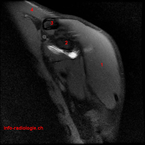
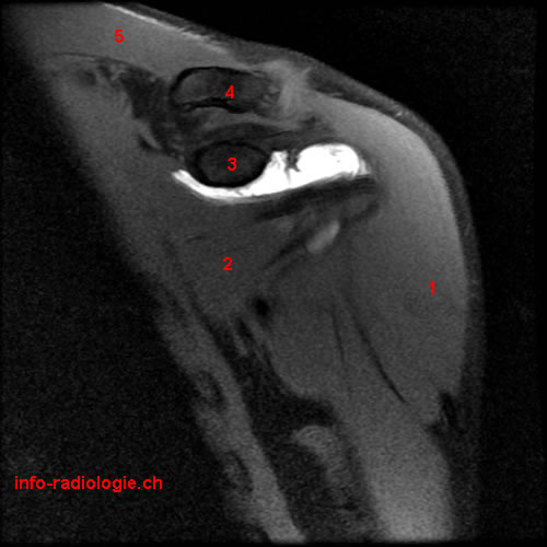
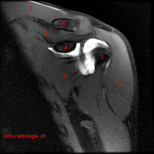
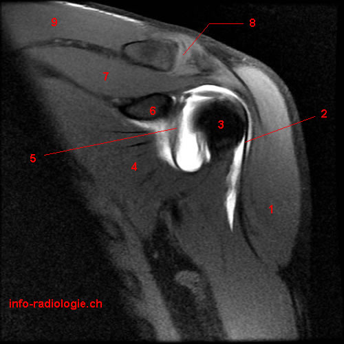
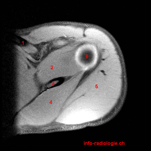
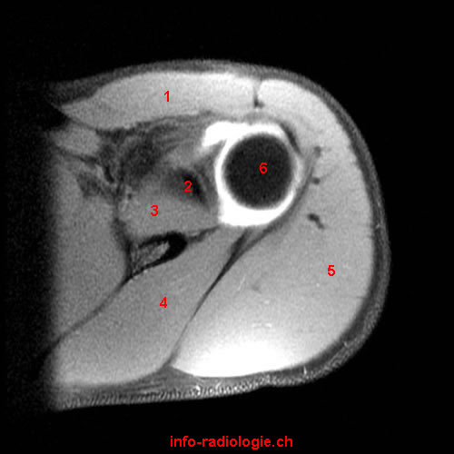
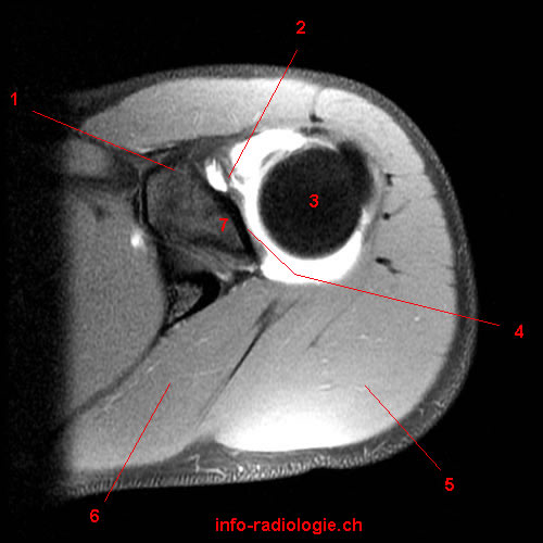
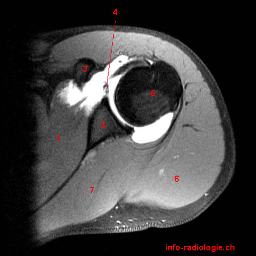
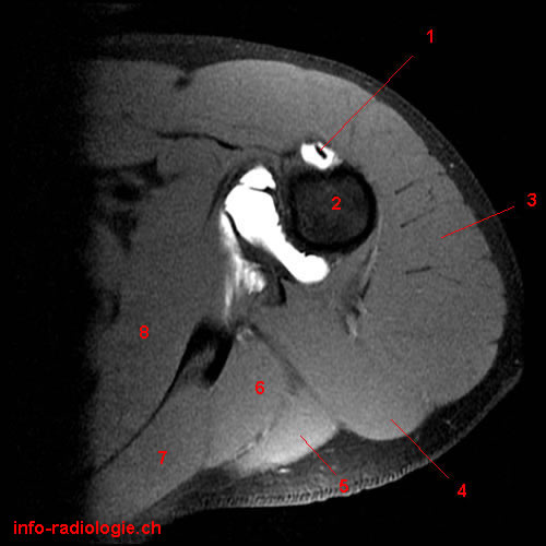
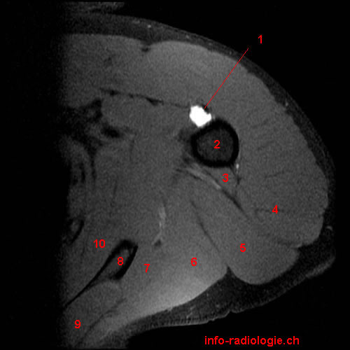
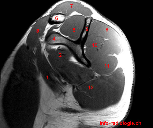
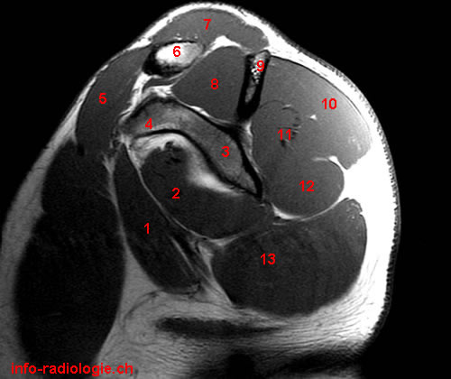
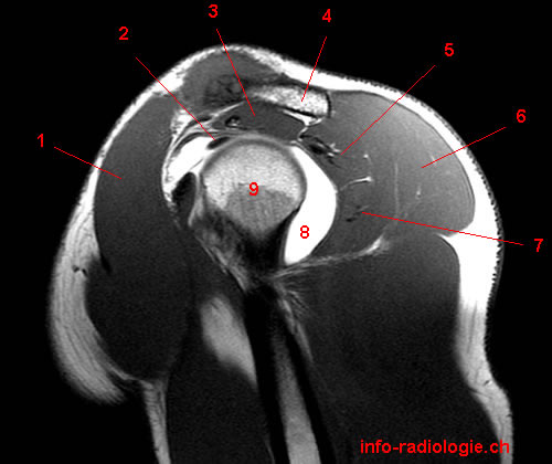
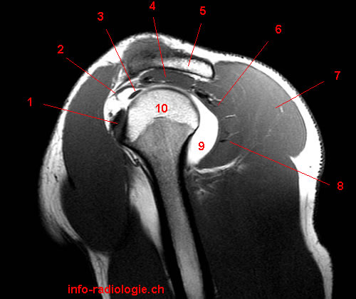
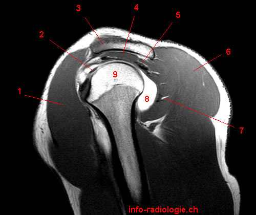
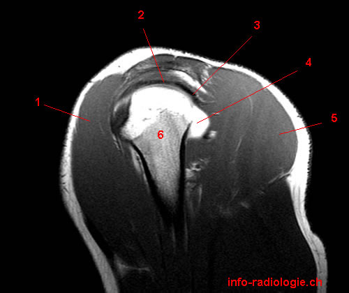
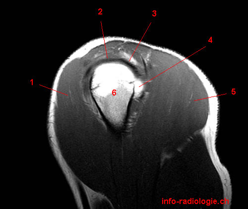
The three bones include the collarbone (clavicle), the shoulder blade (scapula), and the upper arm and bone (humerus)(3). The ends of these bones form the joints of the shoulder girdle.

The shoulder joints, sternoclavicular joints, and acromioclavicular joints facilitate movement(4). The shoulder joints allow forward, circular, and backward movements of the shoulder. 

Meanwhile, the sternoclavicular joints are where the clavicle meets the sternum(5). In the acromioclavicular joints, the clavicle or collarbone meets the acromion (highest point of the shoulder).

Joints, such as the sternoclavicular joints, are bound together by ligaments.

Ligaments are white and flexible bands of fibrous tissues that bind joints together and connect different bones and cartilage(6). These include the joint capsule, ligaments that attach the clavicle to the acromion, and ligaments attached to the coracoid process to connect the clavicle to the scapula.

Tendons refer to the tough cords of tissue that connect the muscles to bones(7).

The shoulder’s intrinsic muscles include the deltoid, teres major, and supraspinatus or rotator cuff(8). The deltoid’s anterior aspect is involved in flexion and medial rotation of the arm.

The middle aspect is involved in the abduction of the arm. Meanwhile, the posterior aspect manages the extension and lateral rotation of the arm.

The teres major is responsible for the medial rotation of the arm(9). The rotator cuff helps initiate arm abduction and stabilizes glenohumeral joints. 

Glenohumeral joints are formed where a ball (head) at the top of the humerus fits into a shallow socket (glenoid) in the scapula, facilitating various movements(10).

Despite its flexibility, the shoulder is prone to injury because the head of the upper arm is bigger than the socket of the shoulder(11). Moreover, identifying the cause of shoulder pain may be challenging(12).

Other shoulder problems include dislocation, separation, impingement syndrome, and bursitis(13). Impingement syndrome results from the excessive rubbing of the rotator cuff and shoulder blade.

Meanwhile, bursitis occurs when tendonitis and impingement syndrome cause inflammation of the bursa sacs protecting the shoulder(14)

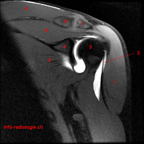
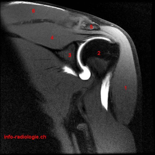
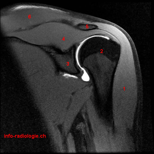
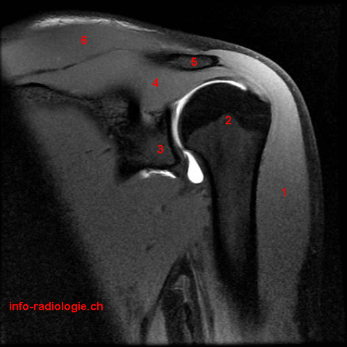
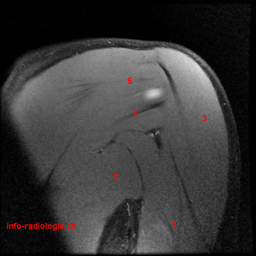
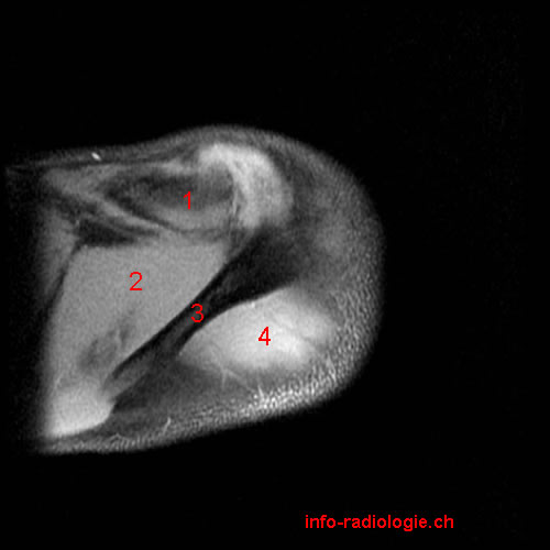
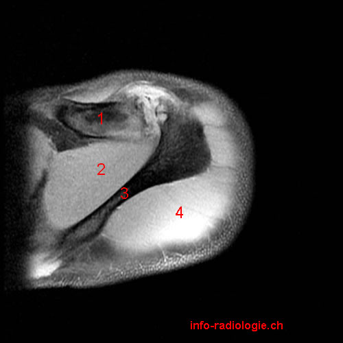
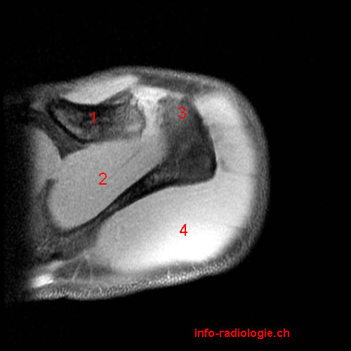
MR Arthrography of the Shoulder

Shoulder pain is a common condition related to the musculoskeletal system(15). If not properly diagnosed, shoulder pain can lead to functional disability. Thus, it is vital to identify the right diagnostic tool to use.

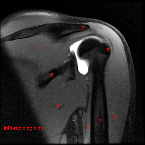
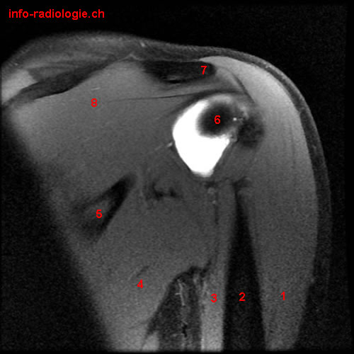
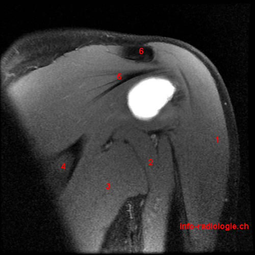
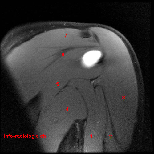
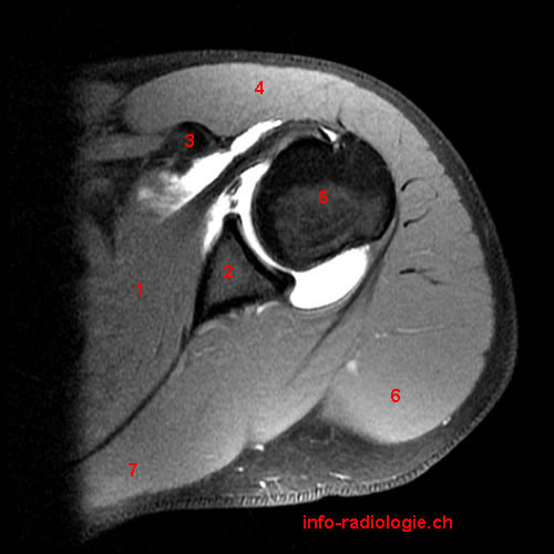
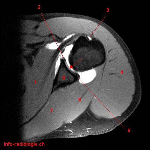
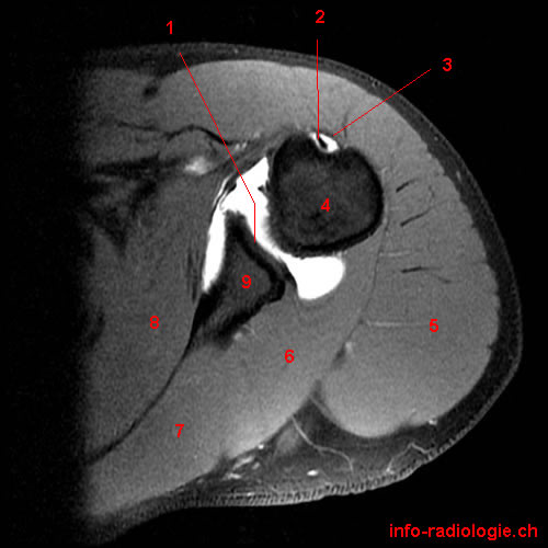
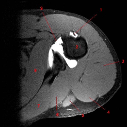
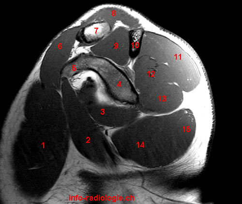
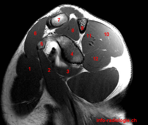
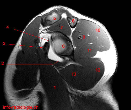
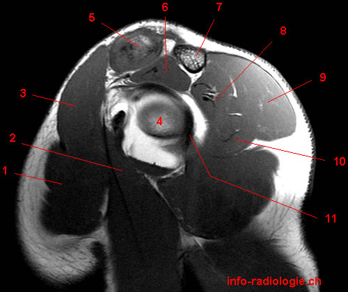
One of these tools is magnetic resonance (MR) arthrography. It has high accuracy, especially in assessing glenohumeral instability(16). The procedure provides superior detailing of cartilaginous, ligamentous, and labral structures. 

During an MR arthrography, the joint space is accessed by guided puncture followed by an injection of a contrast agent, making the procedure invasive(17). A contrast agent is a special dye used to enhance image quality(18).

Some people may think that MR arthrography is a painful procedure. However, a 2018 study published in Radiologia Brasileira showed that this method is less painful than patients think(19).

What to Expect From a Shoulder MR Arthrography

Before the Procedure

The physician may provide restrictions on eating or drinking before the exam. Patients must remove metal objects, like jewelry, hearing aids, and eyeglasses, to ensure that these do not interfere with the imaging procedure.

Moreover, the patients must disclose their pregnancy, any allergies, or current medications. Lactating women should also inform the doctor to avoid harming the baby.

The doctor may give additional or special instructions based on the patient’s needs.

During the Procedure

MR arthrography has two parts. During the first part, the patient lies on an X-ray fluoroscopy table.

The radiologist looks at the examined area via fluoroscopy and marks the joint space with a felt pen. This doctor then cleans the skin and numbs the area with a local anesthetic. 

To minimize discomfort at the beginning of the procedure, the radiologist injects a local anesthetic, signaled by a sharp burning sensation. 

The radiologist places a thin needle into the symptomatic joint area and injects contrast material. This contrast material helps improve the quality of the images(20)

The second part of MR arthrography is magnetic resonance imaging (MRI). The patient lies on a movable scanning table with the technologist placing the patient into position. This procedure requires the patient to stay still to avoid obtaining blurred images.

After the Procedure

The radiologist interprets the results and submits a report to the referring physician. In turn, the physician shares the results with the patient.

Unless the doctor provides special instructions, the patient may return to their regular diet and prescribed medications after the procedure.


  1. InformedHealth.org [Internet]. Cologne, Germany: Institute for Quality and Efficiency in Health Care (IQWiG); 2006-. How does the shoulder work? 2020 Feb 13. Available from: https://www.ncbi.nlm.nih.gov/books/NBK554696/ 
  2. Ibid. 
  3. Ibid. 
  4. Johns Hopkins and Medicine. Shoulder Pain and Problems. Retrieved from https://www.hopkinsmedicine.org/health/conditions-and-diseases/shoulder-pain-and-problems 
  5. Ibid. 
  6. Ibid.
  7. Ibid.
  8. Miniato MA, Anand P, Varacallo M. Anatomy, Shoulder and Upper Limb, Shoulder. [Updated 2020 Jul 31]. In: StatPearls [Internet]. Treasure Island (FL): StatPearls Publishing; 2020 Jan-. Available from: https://www.ncbi.nlm.nih.gov/books/NBK536933/ 
  9. Ibid.
  10. Arthritis Foundation. Shoulder Anatomy. Retrieved from https://www.arthritis.org/health-wellness/about-arthritis/where-it-hurts/shoulder-anatomy 
  11. Johns Hopkins and Medicine. Op Cit.
  12. InformedHealth.org [Internet]. (2006). Op Cit.
  13. Johns Hopkins and Medicine. Op Cit.
  14. Ibid.
  15. do Nascimento, P., Amaral, A. M., & de Almeida, J. (2018). Magnetic resonance arthrography of the shoulder: a painful procedure? Radiologia brasileira, 51(2), 81–86. doi.org/10.1590/0100-3984.2016.0226. https://www.ncbi.nlm.nih.gov/pmc/articles/PMC5935400/ 
  16. Ibid.
  17. Ibid.
  18. Cedars Sinai. MRI & MRA Procedures. Retrieved from https://www.cedars-sinai.org/programs/heart/clinical/aortic/diagnostic-tests/mri-mra.html 
  19. do Nascimento, P. (2018). Op Cit.
  20. Ibid

Need more?