The wrist consists of multiple joints where the bones of the arm and hand meet to facilitate movement(1). Research showed that magnetic resonance imaging (MRI) of the wrist helps influence clinicians’ diagnoses and management plans(2).
MRI is a non-invasive imaging technology that produces detailed images of body tissues or organs to detect a disease or monitor the body’s reaction to a specific treatment(3). MRI has been used to diagnose wrist and hand diseases, like rheumatoid arthritis, osteoarthritis, and ligamentous or tendinous injuries(4).
Thus, it is essential to understand wrist anatomy and see how MRI can help diagnose wrist-related conditions.
How Does Wrist MRI Work?
The wrist is one of the most complex joints in the human body(5). Like other joints, the wrist is supported by several ligaments and tendons that provide structural stability and movement.
Degeneration of any of the structures may lead to various types of pain and dysfunction, which may not be easily diagnosed because of the wrist’s complex anatomy.
MRI helps assess the wrist with high-resolution, multiplanar imaging with no ionizing radiation(6). Thus, it is an ideal investigation tool in assessing pathologic conditions related to joints.
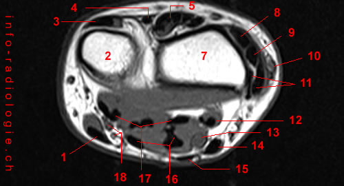
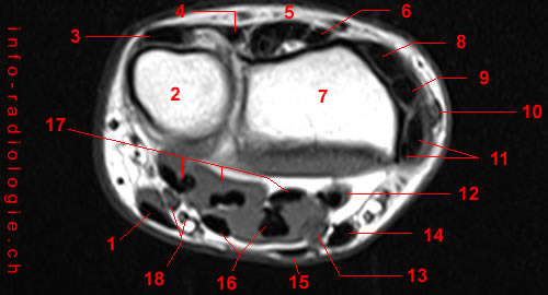
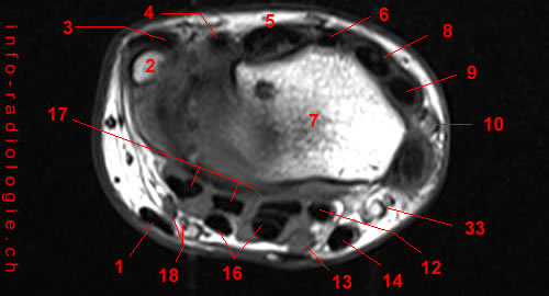
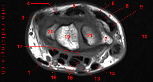
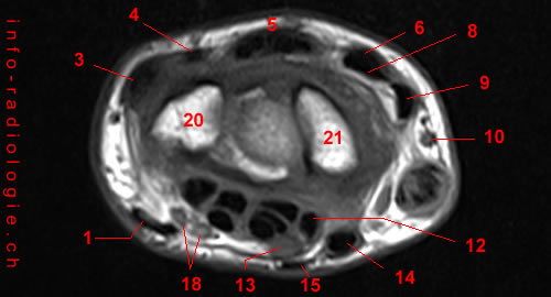
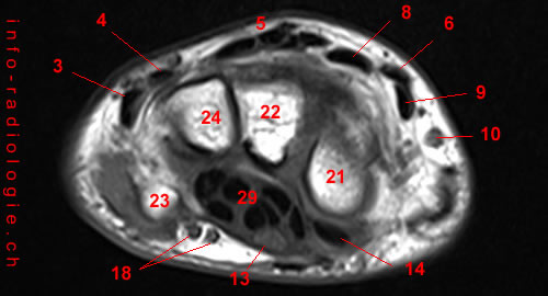
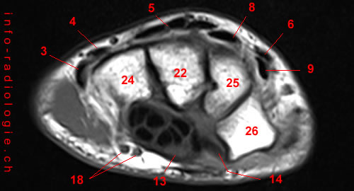
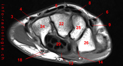
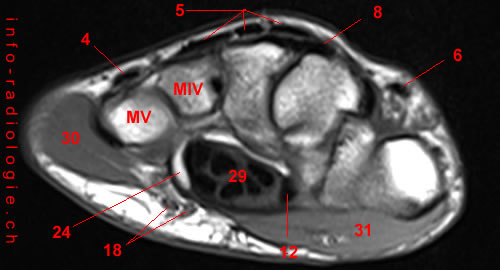
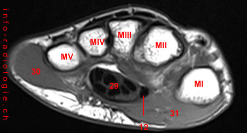
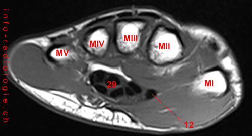
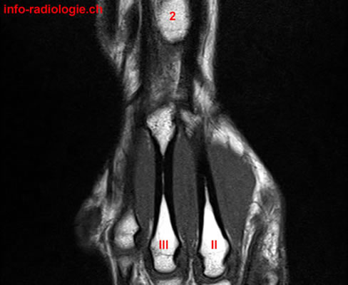
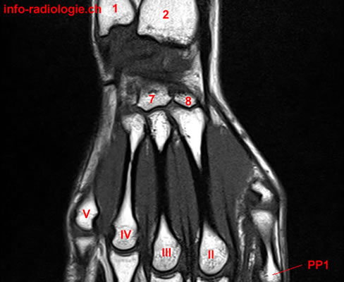
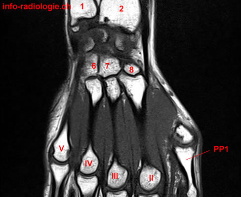
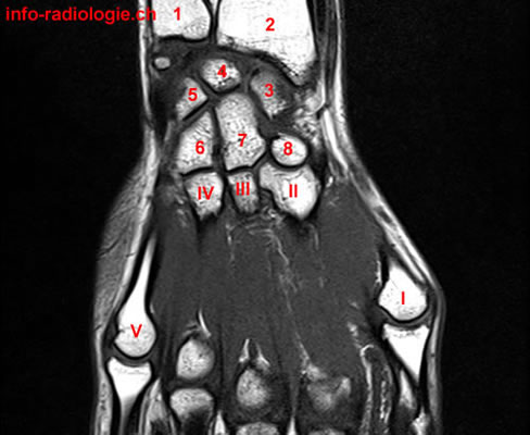
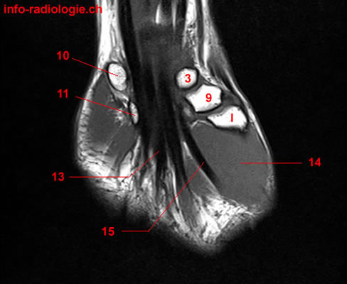
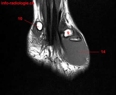
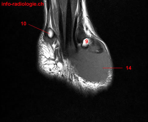
MRI images can be obtained in coronal, axial, and sagittal views(7). Axial views feature tendons, blood vessels, nerves, and the two passageways of the radiocarpal joint or wrist (carpal tunnel and ulnar canal).
Moreover, the bones and ligaments are visible in axial views. However, coronal views may produce a clearer assessment.
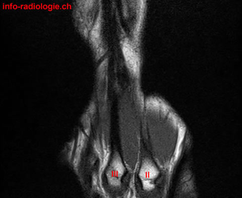
Meanwhile, sagittal views emphasize the carpal bone alignment. They are often optional as sagittal views offer limited benefits in understanding wrist anatomy.
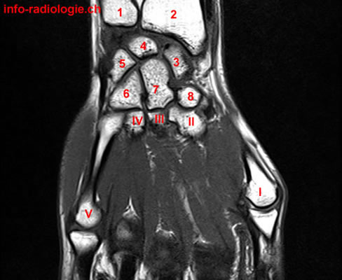
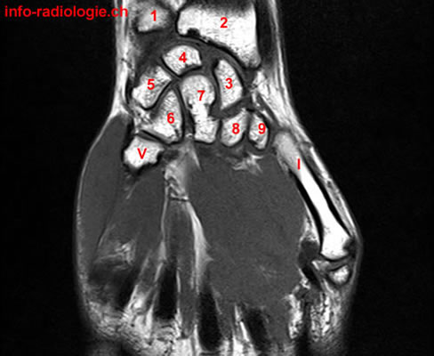
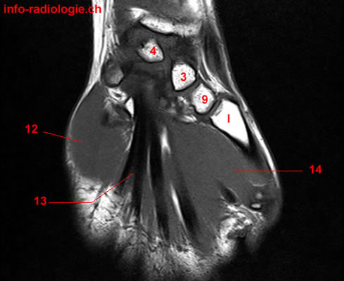
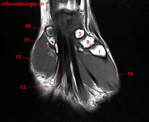
Bones
MRI of the wrist includes assessing the wrist’s bony structures, the captured distal radius and ulna to the bases, and proximal parts of the metacarpals (long bones within the hand), including the proximal and distal row of the wrist (carpal) bones(8).
Fractures, dislocations, scapholunate advanced collapse (SLAC), and scaphoid nonunion advanced collapse (SNAC) may be detected.
SLAC is a wrist malalignment associated with post-traumatic or spontaneous osteoarthritis of the wrist(9). This condition may occur with undiagnosed or untreated scapholunate dissociation (disruption of scaphoid and lunate).
Meanwhile, SNAC occurs with scaphoid fractures, which eventually results in the nonunion of these fractures(10).
Occult fractures show a band of low signal intensity along the scaphoid waist with the absence of a clear fracture line(11). Typical areas of bone contusion include the scaphoid, the tubercle of the trapezium (radial-sided pain), the distal radius, and the hook of the hamate (ulnar or volar-sided pain).
Ligaments
Imaging wrist ligaments is usually tricky as they are thin with an oblique course(12). These wrist ligaments have two types: intrinsic and extrinsic ligaments.
Intrinsic ligaments attach to carpal bones independently, while extrinsic ligaments connect the radius, ulna, or metacarpals to the carpal bones. Both ligaments are crucial for carpal stability, with intrinsic ligaments as the main stabilizer.
The most relevant intrinsic ligament of the wrist that can be examined with MRI is the scapholunate ligament (SLL)(13). The SLL consists of three bands: dorsal, volar, and central bands. Dorsal bands can be easily viewed on axial and coronal images.
Meanwhile, volar bands are less prone to injury as they are protected by their adjacent, strong extrinsic ligament. The central band is usually perforated in adults without clear consequence for wrist biomechanics.
MRI helps assess the presence of scapholunate ligament injury (SLL)(14). Axial images show the dorsal component as a thick, band-like structure with low signal intensity, while the volar component is heterogeneous.
MRI may also show irregular morphology of SSL, abnormal signal intensity, and fluid transecting the ligaments.
Triangular Fibrocartilage Complex (TFCC)
The triangular fibrocartilage complex (TFCC) is an essential structure in the wrist that consists of tough fibrous tissues and cartilage(15). Moreover, the TFCC serves as a stabilizer, supporting the joints between the end of the forearm bones (radius and ulna).
Types of TFCC tears may include natural wear and tears from injury, which may come from a fall on the hand or wrist or a fracture at the end of the radius.
TFCC injuries may appear in MRI as degenerative signal changes, non-communicating defects (or partial tears) of the surfaces, and communicating defects (full-thickness tears)(16).
Some people may not feel pain or instability from TFCC tears(17). MRI studies often observe tears in individuals who do not feel pain or encounter problems using the wrist(18).
Moreover, the prevalence of these TFCC tears may increase as patients age(19).
Tendons
Tendons of the wrist have two subgroups: flexor tendons and extensor tendons(20). Both demonstrate the typical low signal intensity and constant diameter on all sequences. In MRI, tendinopathy appears as signal and thickness changes, which may lead to the partial or full thickness of tears.
Moreover, tendons of the wrist are surrounded by a synovial sheath. Hence, they may be at risk for synovium-based inflammatory diseases, including rheumatoid arthritis(21).
During an MRI, tendon involvement in rheumatoid arthritis may manifest with synovial hypertrophy and soft-tissue thickening, inflammatory changes, and the presence of tendon sheath fluid.
Thus, while the wrist is one of the most complex joints, MRI can help assess wrist anatomy and detect conditions associated with the wrist.