This webpage presents the anatomical structures found on ankle CT.
Scout view (coronal reconstructions).
-
Scout view (coronal reconstructions).
-
CT-scan of the ankle (coronal reconstruction). Image 2. 1, Fibula. 2, Tibia. 3, Talus.
-
CT-scan of the ankle (coronal reconstruction). Image 3. 1, Fibula. 2, Tibia. 3, Talus.
-
CT-scan of the ankle (coronal reconstruction). Image 4. 1, Fibula. 2, Tibia. 3, Talus.
-
CT-scan of the ankle (coronal reconstruction). Image 5. 1, Fibula. 2, Tibia. 3, Talus.
-
CT-scan of the ankle (coronal reconstruction). Image 6. 1, Fibula. 2, Tibia. 3, Talus. 4, Navicular.
-
CT-scan of the ankle (coronal reconstruction). Image 7. 1, Fibula. 2, Tibia. 3, Talus. 4, Navicular. 5, Intermediate cuneiform.
-
CT-scan of the ankle (coronal reconstruction). Image 8. 1, Fibula. 2, Tibia. 3, Talus. 4, Navicular. 5, Intermediate cuneiform. 6, Medial cuneiform. 7, Base of 2nd metatarsal. 8, Lateral cuneiform.
-
CT-scan of the ankle (coronal reconstruction). Image 9. 1, Fibula. 2, Tibia. 3, Talus. 4, Navicular. 5, Intermediate cuneiform. 6, Medial cuneiform. 7, Base of 2nd metatarsal. 8, Lateral cuneiform.
-
CT-scan of the ankle (coronal reconstruction). Image 10. 1, Fibula. 2, Tibia. 3, Talus. 4, Navicular. 5, Medial cuneiform. 6, Base of 1st metatarsal. 7, Base of 2nd metatarsal. 8, Base of 3rd metatarsal. 9, Lateral cuneiform. 10, Cuboid. 11, Calcaneus.
-
CT-scan of the ankle (coronal reconstruction). Image 11. 1, Fibula. 2, Tibia. 3, Talus. 4, Navicular. 5, Medial cuneiform. 6, Base of 1st metatarsal. 7, Base of 2nd metatarsal. 8, Base of 3rd metatarsal. 9, Lateral cuneiform. 10, Cuboid. 11, Calcaneus.
-
CT-scan of the ankle (coronal reconstruction). Image 12. 1, Calcaneus. 2, Talus. 3, Navicular. 4, Medial cuneiform. 5, Base of 1st metatarsal. 6, 2nd metatarsal. 7, 3rd metatarsal. 8, Base of 4th metatarsal. 9, Cuboid.
-
CT-scan of the ankle (coronal reconstruction). Image 13. 1, Calcaneus. 2, Talus. 3, Navicular. 4, Medial cuneiform. 5, Base of 1st metatarsal. 6, 2nd metatarsal. 7, 2nd proximal phalanx. 8, 2nd middle phalanx. 9, 2nd distal phalanx. 10, 5th metatarsophalangeal joint. 11, Cuboid.
-
CT-scan of the ankle (coronal reconstruction). Image 14. 1, Base of 5th proximal phalanx. 2, 5th metatarsal. 3, Cuboid. 4, Calcaneus. 5, Navicular. 6, Medial cuneiform. 7, Base of 1st metatarsal. 8, 2nd metatarsal.
-
CT-scan of the ankle (coronal reconstruction). Image 15. 1, 5th metatarsal. 2, Cuboid. 3, Calcaneus. 4, Navicular. 5, Medial cuneiform. 6, 1st metatarsal. 7, 2nd metatarsophalangeal joint. 8, Distal phalanx. 9, base of 3rd proximal phalanx. 10, base of 4th proximal phalanx.
-
CT-scan of the ankle (coronal reconstruction). Image 16. 1, Base of 5th metatarsal. 2, Cuboid. 3, Calcaneus. 4, Navicular. 5, Medial cuneiform. 6, 1st metatarsal. 7, 1st proximal phalanx. 8, 1st distal phalanx. 9, base of 2nd proximal phalanx. 10, 4th metatarsal.
-
CT-scan of the ankle (coronal reconstruction). Image 17. 1, Base of 5th metatarsal. 2, Calcaneus. 3, Medial cuneiform. 4, 1st metatarsal. 5, 1st metatarsophalangeal joint. 6, 1st distal phalanx.
-
CT-scan of the ankle (coronal reconstruction). Image 18. 1, Base of 5th metatarsal. 2, Cuboid. 3, Calcaneus. 4, Medial cuneiform. 5, 1st metatarsal. 6, 1st proximal phalanx. 7, 1st distal phalanx.
-
CT-scan of the ankle (coronal reconstruction). Image 19. 1, Calcaneus. 2, 1st metatarsophalangeal joint.
-
CT-scan of the ankle (coronal reconstruction). Image 20. 1, Calcaneus. 2, 1st metatarsal. 3, 1st proximal phalanx.
-
Scout view (Axial reconstruction)
-
CT-scan of the ankle (axial reconstruction). Image 1. 1, Fibula. 2, Tibia.
-
CT-scan of the ankle (axial reconstruction). Image 2. 1, Fibula. 2, Tibia.
-
CT-scan of the ankle (axial reconstruction). Image 3. 1, Fibula. 2, Tibia. 3, Calcaneus.
-
CT-scan of the ankle (axial reconstruction). Image 4. 1, Fibula. 2, Tibia. 3, Calcaneus.
-
CT-scan of the ankle (axial reconstruction). Image 5. 1, Fibula. 2, Tibia. 3, Talus. 4, Calcaneus.
-
CT-scan of the ankle (axial reconstruction). Image 6. 1, Fibula. 2, Tibia. 3, Talus. 4, Calcaneus.
-
CT-scan of the ankle (axial reconstruction). Image 7. 1, Fibula. 2, Tibia. 3, Talus. 4, Calcaneus.
-
CT-scan of the ankle (axial reconstruction). Image 8. 1, Fibula. 2, Tibia (medial malleolus). 3, Talus. 4, Calcaneus.
-
CT-scan of the ankle (axial reconstruction). Image 9. 1, Fibula. 2, Talus. 3, Tibia (medial malleolus). 4, Calcaneus.
-
CT-scan of the ankle (axial reconstruction). Image 10. 1, Talus. 2, Calcaneus.
-
CT-scan of the ankle (axial reconstruction). Image 11. 1, Talus. 2, Calcaneus.
-
CT-scan of the ankle (axial reconstruction). Image 12. 1, Talus. 2, Calcaneus.
-
CT-scan of the ankle (axial reconstruction). Image 13. 1, Calcaneus. 2, Talus.
-
CT-scan of the ankle (axial reconstruction). Image 14. 1, Calcaneus. 2, Talus. 3, Navicular. 4, Cuboid.
-
CT-scan of the ankle (axial reconstruction). Image 15. 1, Cuboid 2, Navicular.
-
CT-scan of the ankle (axial reconstruction). Image 16. 1, Cuboid 2, Navicular.
-
CT-scan of the ankle (axial reconstruction). Image 17. 1, Cuboid. 2, Lateral cuneiform. 3, Navicular. 4, Medial cuneiform.
-
CT-scan of the ankle (axial reconstruction). Image 18. 1, 5th metatarsal. 2, Cuboid. 3, Lateral cuneiform. 4, Intermediate cuneiform. 5, Medial cuneiform.
-
CT-scan of the ankle (axial reconstruction). Image 19. 1, 5th metatarsal. 2, Cuboid. 3, Lateral cuneiform. 4, Intermediate cuneiform. 5, Medial cuneiform.
-
CT-scan of the ankle (axial reconstruction). Image 20. 1, 5th metatarsal. 2, 4th metatarsal. 3, Lateral cuneiform. 4, Intermediate cuneiform. 5, Medial cuneiform. 6, 2nd metatarsal. 7, 3rd metatarsal.
-
CT-scan of the ankle (axial reconstruction). Image 21. 1, 5th metatarsal. 2, 4th metatarsal. 3, 3rd metatarsal. 4, 2nd metatarsal. 5, Medial cuneiform. 6, 1st metatarsal.
-
CT-scan of the ankle (axial reconstruction). Image 22. 1, 5th metatarsal. 2, 4th metatarsal. 3, 3rd metatarsal. 4, 2nd metatarsal. 5, 1st metatarsal.
-
CT-scan of the ankle (axial reconstruction). Image 23. 1, 5th metatarsal. 2, 4th metatarsal. 3, 3rd metatarsal. 4, 2nd metatarsal. 5, 1st metatarsal.
-
Scout view (sagittal reconstruction)
-
CT-scan of the ankle (sagittal reconstruction). Image 2. 1, Tibia (medial malleolus). 2, 1st Metatarsophalangeal joint.
-
CT-scan of the ankle (sagittal reconstruction). Image 3. 1, Tibia (medial malleolus). 2, Navicular. 3, 1st metatarsal. 4, Proximal phalanx.
-
CT-scan of the ankle (sagittal reconstruction). Image 4. 1, Tibia. 2, Navicular. 3, 1st metatarsal. 4, Distal phalanx. 5, Proximal phalanx. 6, Medial cuneiform. 7, Calcaneus. 8, Talus.
-
CT-scan of the ankle (sagittal reconstruction). Image 5. 1, Tibia. 2, Talus. 3, Medial cuneiform. 4, Proximal phalanx. 5, Distal phalanx. 6, 1st metatarsal. 7, Navicular. 8, Calcaneus.
-
CT-scan of the ankle (sagittal reconstruction). Image 6. 1, Tibia. 2, Talus. 3, Medial cuneiform. 4, Proximal phalanx. 5, Distal phalanx. 6, 1st metatarsal. 7, Navicular. 8, Calcaneus.
-
CT-scan of the ankle (sagittal reconstruction). Image 7. 1, Tibia. 2, Talus. 3, Navicular. 4, Medial cuneiform. 5, Base of 1st metatarsal. 6, 1st distal phalanx. 7, Calcaneus.
-
CT-scan of the ankle (sagittal reconstruction). Image 8. 1, Tibia. 2, Talus. 3, Navicular. 4, Medial cuneiform. 5, Base of 1st metatarsal. 6, Calcaneus.
-
CT-scan of the ankle (sagittal reconstruction). Image 9. 1, Tibia. 2, Talus. 3, Navicular. 4, Medial cuneiform. 5, Base of 1st metatarsal. 6, 2nd metatarsal. 7, 2nd proximal phalanx. 8, Intermediate cuneiform. 9, Calcaneus.
-
CT-scan of the ankle (sagittal reconstruction). Image 10. 1, Tibia. 2, Talus. 3, Navicular. 4, Intermediate cuneiform. 5, 2nd metatarsal. 6, Metatarsophalangeal joint. 7, Distal interphalangeal joint. 8, Proximal interphalangeal joint. 9, Cuboid. 10, Calcaneus.
-
CT-scan of the ankle (sagittal reconstruction). Image 11. 1, Tibia. 2, Talus. 3, Navicular. 4, Intermediate cuneiform. 5, 2nd metatarsal. 6, 2nd Middle phalanx. 7, Lateral cuneiform. 8, Cuboid. 9, Calcaneus.
-
CT-scan of the ankle (sagittal reconstruction). Image 12. 1, Tibia. 2, Talus. 3, Navicular. 4, Intermediate cuneiform. 5, base of 2nd metatarsal. 6, 3rd metatarsal. 7, base of 3rd metatarsal. 8, Lateral cuneiform. 9, Cuboid. 10, Calcaneus. 11, Fibula.
-
CT-scan of the ankle (sagittal reconstruction). Image 13. 1, Tibia. 2, Talus. 3, Lateral cuneiform. 4, 3rd metatarsal. 5, 3rd Metatarsophalangeal joint. 6, Cuboid. 7, Calcaneus. 8, Fibula.
-
CT-scan of the ankle (sagittal reconstruction). Image 14. 1, Fibula. 2, Cuboid. 3, Lateral cuneiform. 4, Base of 3rd metatarsal. 5, 3rd proximal interphalangeal joint. 6, base of 4th metatarsal. 7, Calcaneus.
-
CT-scan of the ankle (sagittal reconstruction). Image 15. 1, Fibula. 2, Cuboid. 3, Lateral cuneiform. 4, Base du 3rd metatarsal. 5, 3rd distal interphalangeal joint. 6, 4th metatarsal. 7, Calcaneus.
-
CT-scan of the ankle (sagittal reconstruction). Image 16. 1, Fibula. 2, Cuboid. 3, 4th metatarsal. 4, 4th Metatarsophalangeal joint. 5, base of 5th metatarsal.
-
CT-scan of the ankle (sagittal reconstruction). Image 17. 1, Cuboid. 2, Base of 4th metatarsal. 3, 4th Proximal interphalangeal joint. 4, 4th Distal phalanx. 5, base of 5th metatarsal.
-
CT-scan of the ankle (sagittal reconstruction). Image 18. 1, 5th metatarsal. 2, 4th Proximal interphalangeal joint. 3, 4th Distal interphalangeal joint.
-
CT-scan of the ankle (sagittal reconstruction). Image 19. 1, 5th metatarsal.
-
CT-scan of the ankle (sagittal reconstruction). Image 20. 1, 5th Metatarsophalangeal joint. 2, Distal phalanx.
1, Fibula. 2, Tibia. 3, Talus. 4, Navicular. 5, Medial cuneiform. 6, Base of 1st metatarsal. 7, Base of 2nd metatarsal. 8, Base of 3rd metatarsal. 9, Lateral cuneiform. 10, Cuboid. 11, Calcaneus.">
1, Calcaneus. 2, Talus. 3, Navicular. 4, Medial cuneiform. 5, Base of 1st metatarsal. 6, 2nd metatarsal. 7, 2nd proximal phalanx. 8, 2nd middle phalanx. 9, 2nd distal phalanx. 10, 5th metatarsophalangeal joint. 11, Cuboid.">
1, 5th metatarsal. 2, Cuboid. 3, Calcaneus. 4, Navicular. 5, Medial cuneiform. 6, 1st metatarsal. 7, 2nd metatarsophalangeal joint. 8, Distal phalanx. 9, base of 3rd proximal phalanx. 10, base of 4th proximal phalanx.">
1, Base of 5th metatarsal. 2, Cuboid. 3, Calcaneus. 4, Navicular. 5, Medial cuneiform. 6, 1st metatarsal. 7, 1st proximal phalanx. 8, 1st distal phalanx. 9, base of 2nd proximal phalanx. 10, 4th metatarsal.">
1, Tibia. 2, Talus. 3, Navicular. 4, Intermediate cuneiform. 5, 2nd metatarsal. 6, Metatarsophalangeal joint. 7, Distal interphalangeal joint. 8, Proximal interphalangeal joint. 9, Cuboid. 10, Calcaneus.">
1, Tibia. 2, Talus. 3, Navicular. 4, Intermediate cuneiform. 5, base of 2nd metatarsal. 6, 3rd metatarsal. 7, base of 3rd metatarsal. 8, Lateral cuneiform. 9, Cuboid. 10, Calcaneus. 11, Fibula.">
Anatomy of the Foot and Ankle
The foot is a structure of the body with numerous joints, bones, muscles, ligaments, and tendons. It is responsible for the coordinated movements of gait and the body’s ability to stand upright(1).
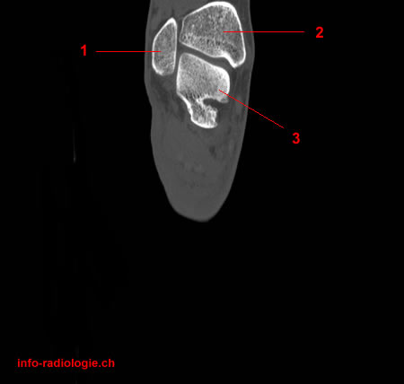
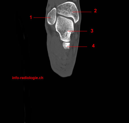
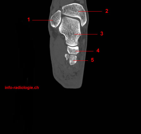
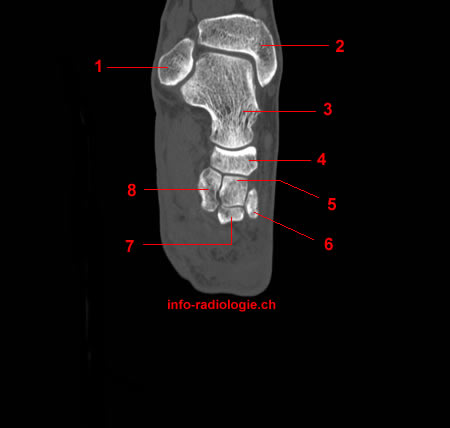
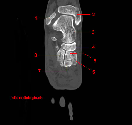
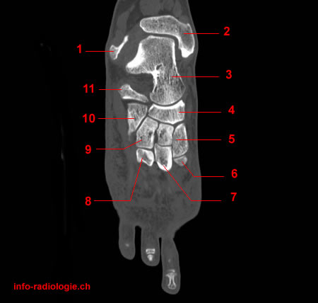
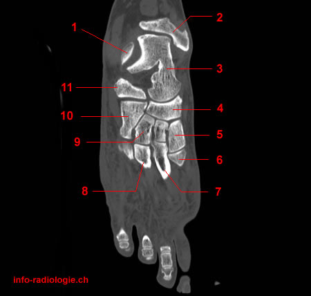
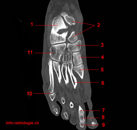
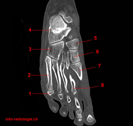
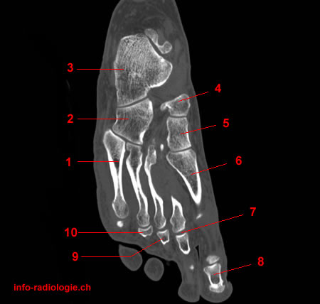
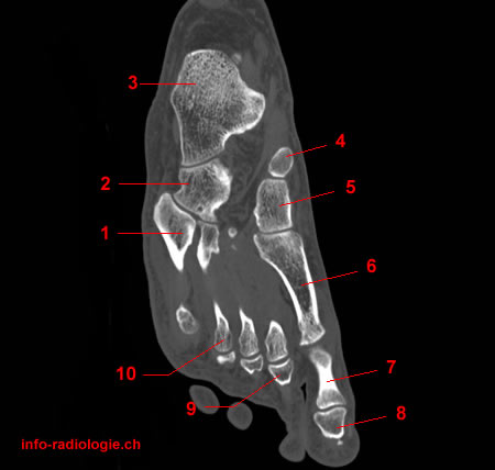
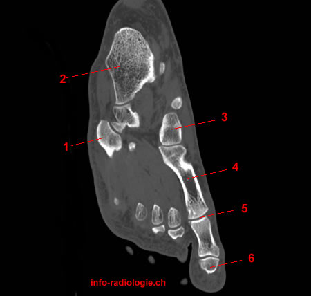
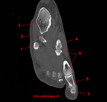
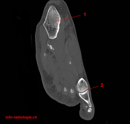
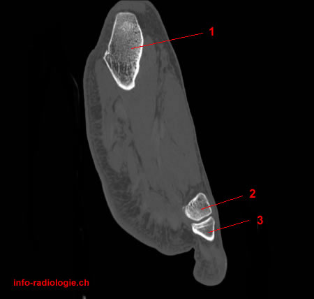
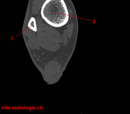
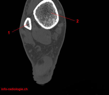
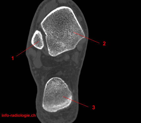
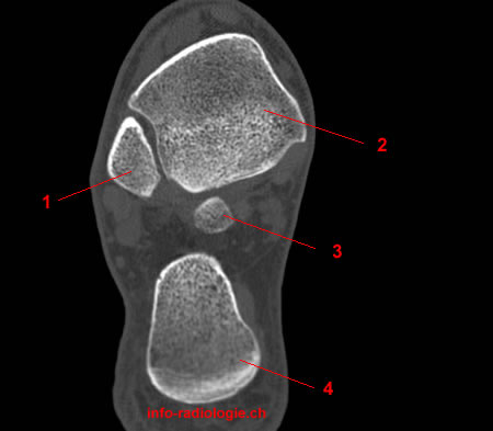
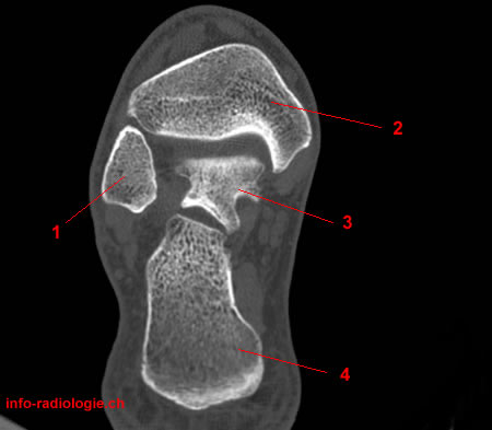
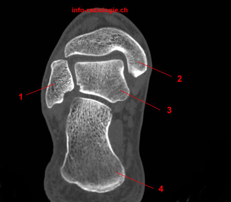
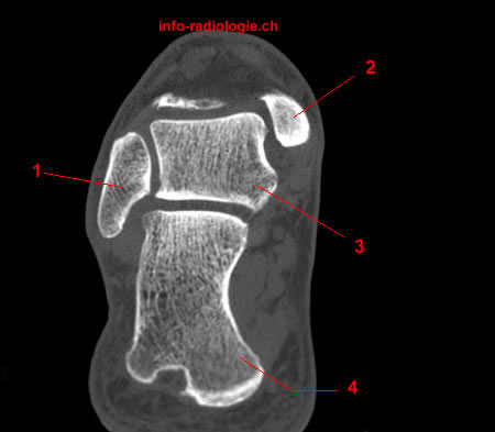
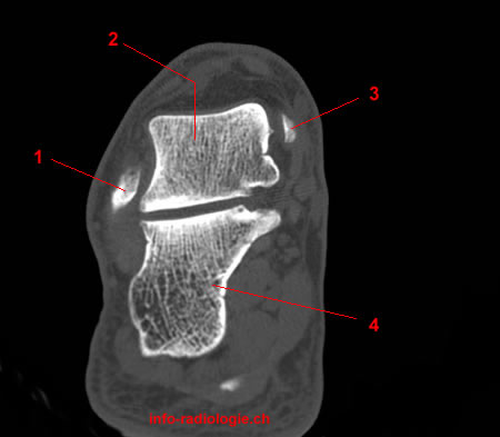
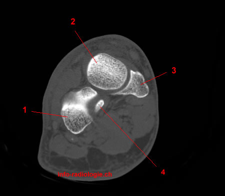
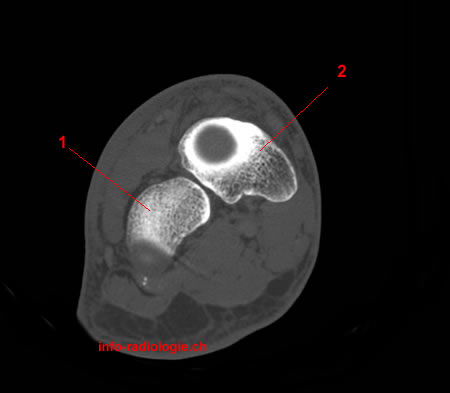
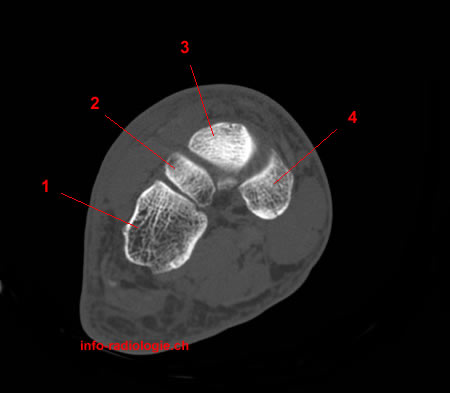
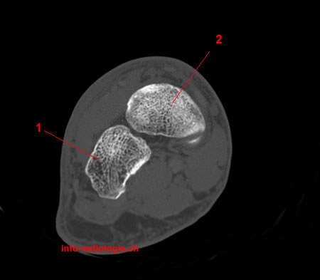
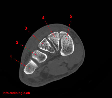
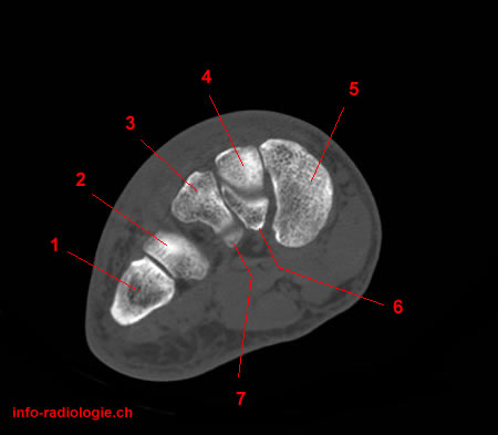
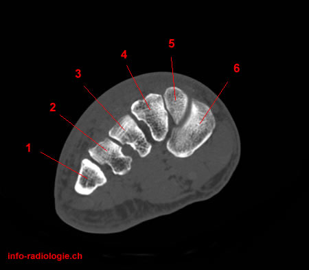
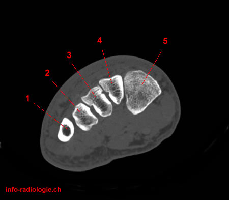
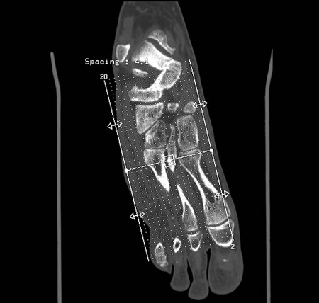
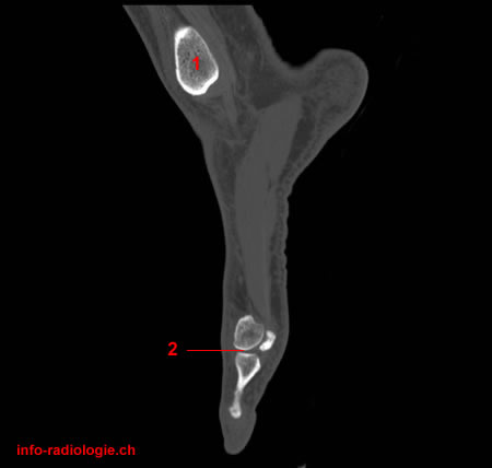
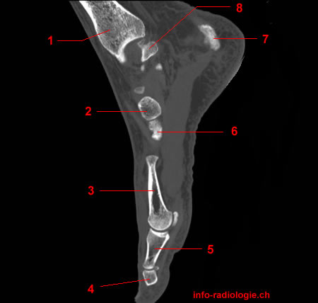
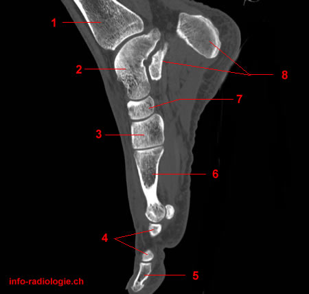
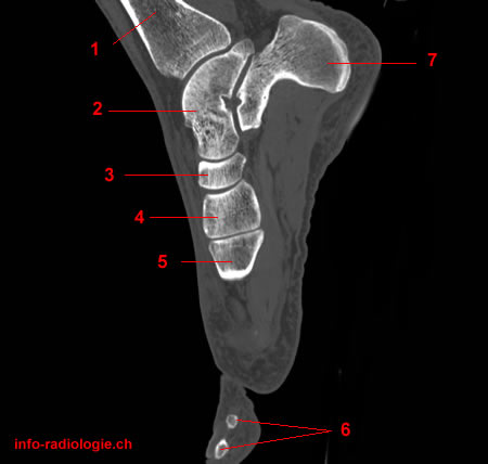
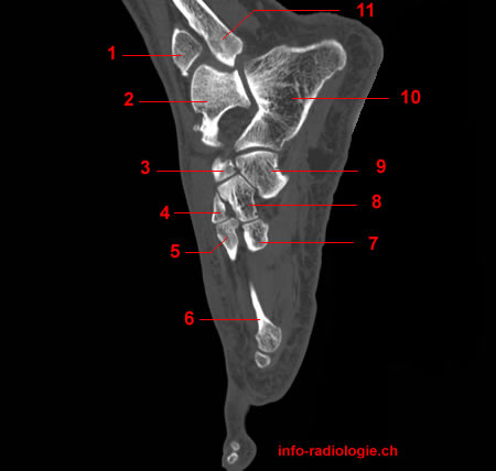
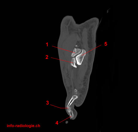
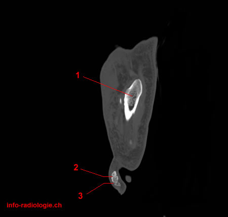
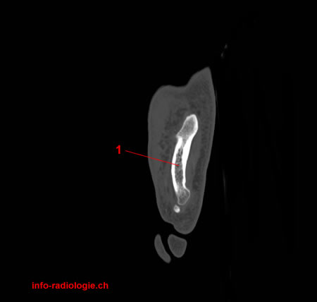
The primary bones in the foot are the tarsals (bones connecting the lower leg to the foot), metatarsals (long bones of the foot), and phalanges (five bones in the toes)(2).
The bones are connected by the tarsometatarsal joints (the joints between the tarsals and metatarsals), midtarsal joint (joints connecting the tarsals and metatarsals), and metatarsophalangeal (MTP) joints (joints between the metatarsals and the phalanges)(3).
Meanwhile, the ankle is a hinge joint found in the talus bone where two bones, tibia and fibula, meet. A strong ligament thickens the ankle on its sides(4).
Ankle and Foot CT
The foot is prone to several types of damage and injuries. Pain and inflammation of the foot may cause limited movement(5).
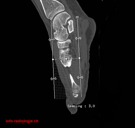
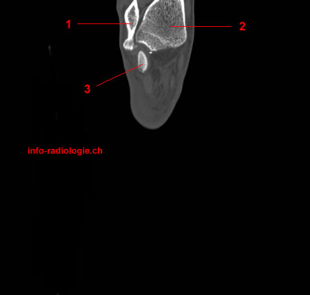
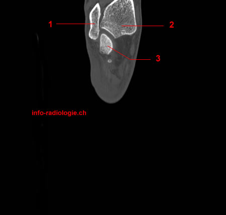
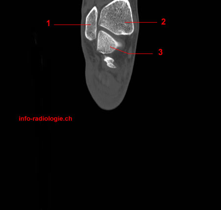
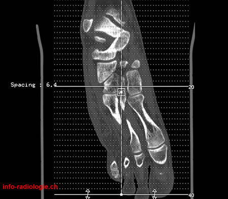
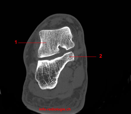
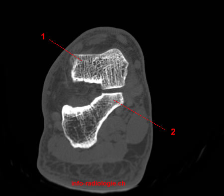
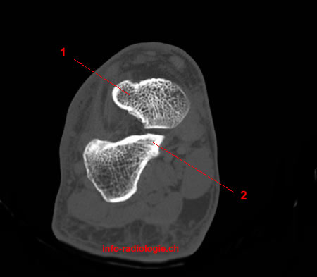
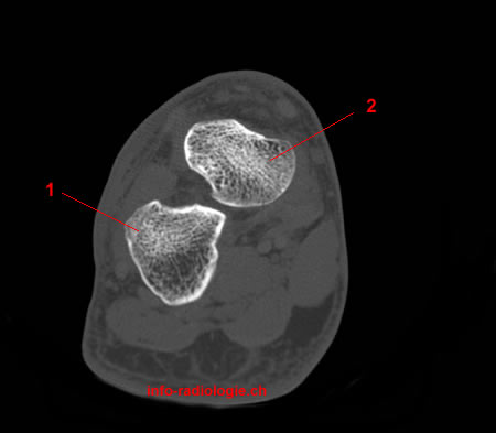
Medical professionals use advanced imaging procedures to diagnose medical problems in the foot. A computed tomography scan of the foot and ankle may help doctors examine any injury or fractures(6).
An ankle and foot computed tomography (CT) may detect the following(7):
- Trauma
- Fractures
- Arthritis
- Osteomyelitis or bone infection
- Foreign bodies
What to Expect From an Ankle and Foot CT
Before the Procedure
The patient must ask the hospital about the individual preparation requirements for their planned ankle and foot CT exam.
Some CT of the feet and ankles may require the injection of intravenous contrast agents. However, the need for a contrast dye depends on the reason for the exam.
If a doctor recommends a CT scan with contrast dye, the patient must submit a recent blood test to assess the function of their kidneys. This requirement should be accomplished before their appointment.
Before the procedure, the patient must also inform their radiologist if they are pregnant.
If a patient suspects that they may be pregnant, then a pregnancy test must be performed before the patient proceeds to take the ankle and foot CT exam.
The patient must also accomplish medical paperwork, such as questionnaires and consent forms, which they must complete upon their arrival at the hospital.
In some hospitals, individuals under the age of 16 must have a consent form signed by a guardian before undergoing the procedure.
Patients should remove all their jewelry and metal items before they enter the room with the CT machine. The items must be removed as they may affect and obscure the images(8).
During the Procedure
If the CT scan includes contrast agents, the doctor may administer the contrast dye before the procedure begins.
The contrast agent highlights the blood vessels in the body during the procedure, allowing the machine to make blood flow more visible in the images(9).
The patient may lie on the CT examination bed, which slides in and out of the CT scanner multiple times.
Once the machine has captured enough images of the ankle and foot, the examination is complete.
A radiologic technologist must always be a short distance away, watching the patient at all times during the procedure.
After the Procedure
After completing a CT examination with intravenous contrast, the patient must flush the contrast dye out of their body by drinking plenty of fluids.
If the patient who took a contrast dye injection is also taking medications for diabetes, the doctor may advise them to stop taking their diabetes medication for two days after the scan.
Stopping the medication may prevent a buildup of the drugs in the patient’s system(10).
A radiologist analyzes the images produced by the CT scan machine. They may draft a report of the results to the referring doctor, who may discuss these findings with the patient.
Risks of a CT Scan
There is a low risk of radiation exposure during the procedure since the CT machine emits a small amount of radiation during a scan.
Patients must tell their physician if they are pregnant or if they suspect they may be pregnant. Exposure to radiation during pregnancy may result in congenital problems(11).
Another risk associated with the procedure is an allergic reaction to the contrast dye. Before the exam, patients must notify their doctor if they are sensitive or allergic to iodine, a chemical present in the contrast dye.
Patients with kidney problems must also notify their physician before the procedure. In some cases, a contrast dye may cause kidney failure(12).
To minimize the risks of ankle and foot CT scan, the patient should discuss any medical concerns with their physician before undergoing the procedure.
- Ficke, J. & Byerly, D. (2020). Anatomy, Bony Pelvis and Lower Limb, Foot. StatPearls. Retrieved from https://www.ncbi.nlm.nih.gov/books/NBK546698/
- Bone And Joint Decade. Global Alliance for Musculoskeletal Health. The Foot & Ankle. Retrieved from http://bjdonline.org/the-foot-ankle/
- Ibid
- O’Rahilly, R., Müller, M., Carpenter, S., & Swenson, R. (2004) BASIC HUMAN ANATOMY A Regional Study of Human Structure. https://www.dartmouth.edu/~humananatomy/part_3/chapter_17.html
- University of Rochester Medical Center. Anatomy of the Foot. Retrieved from https://www.urmc.rochester.edu/encyclopedia/content.aspx?contenttypeid=85&contentid=P07387
- South Western Sydney Local Health District. Foot and Ankle CT. Retrieved from https://www.swslhd.health.nsw.gov.au/medicalImaging/serv_CT_Extremities_Foot.html
- Ibid
- Ibid
- Ibid
- Ibid
- Stanford Healthcare. Risks of Computed Tomography (CT) Scan. Retrieved from https://stanfordhealthcare.org/medical-tests/c/ct-scan/risk-factors.html
- Ibid