Paranasal Sinuses
Paranasal sinuses refer to a group of air-filled spaces around the nasal cavity (a system of air channels that connect the nose with the back of the throat)(1). They facilitate the circulation of the air breathed in and out of the respiratory system(2).
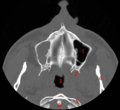
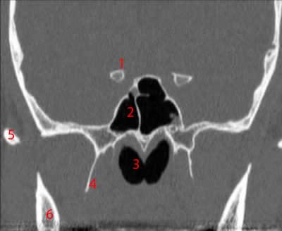
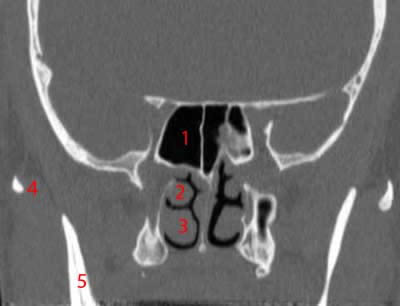
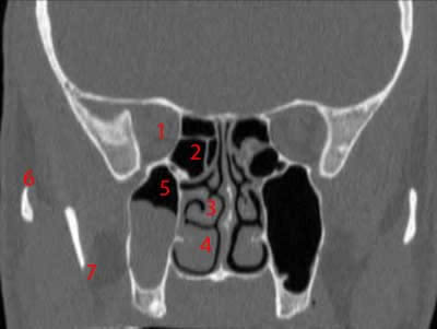
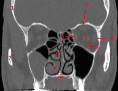
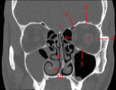
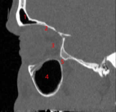
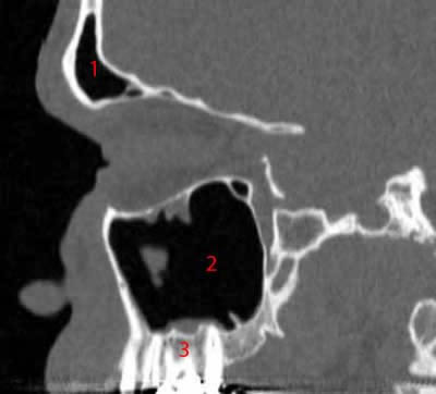
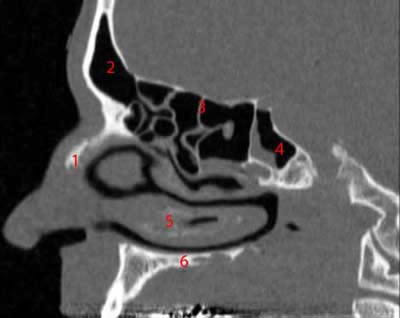
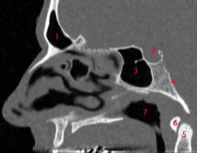
Paranasal sinuses have four different pairs: maxillary sinuses, frontal sinuses, sphenoidal sinuses, and ethmoidal sinuses(3). Their names come from the facial bones where they are located.
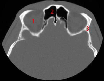
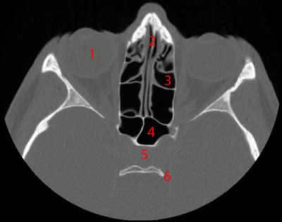
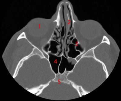
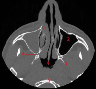
Maxillary sinuses, the largest of all paranasal sinuses, have thin walls usually penetrated by the long roots of the posterior maxillary teeth(4). They primarily lie under the eyes in the maxillary bones.
Meanwhile, frontal sinuses are located superior to the eyes within the frontal lobe(5). Sphenoid sinuses lie within the sphenoid bone.
Various discrete air cells within the ethmoid bone between the nose and the eyes form ethmoidal sinuses(6).
While the function of paranasal sinuses remains unclear, they are associated with the following roles(7):
- Increasing the resonance of the voice
- Decreasing the skull’s relative weight
- Insulating sensitive structures from rapid temperature fluctuations in the nose
- Providing immunological defense
- Humidifying and heating inspired air
- Providing a buffer against facial trauma
Paranasal Sinuses Computed Tomography
A computed tomography (CT) scan combines different X-ray images from various angles around the body(8). It uses computer processing to produce cross-sectional images or slices of the bones, blood vessels, and soft tissues inside the body.
Unlike the typical X-rays, CT scan images produce more detailed images(9). Computed tomography (CT) scans may help examine paranasal sinuses’ anatomy and detect abnormalities(10).
Moreover, a CT scan gives a greater definition of the sinuses(11). It is more sensitive than typical radiography tools in detecting sinus pathology, especially within the sphenoid and ethmoid sinuses.
A CT scan may help detect sinusitis, evaluate sinuses filled with thickened sinus membranes, give additional information on tumors of the nasal cavity, diagnose inflammatory disorders, and plan for surgery by defining anatomy(12).
The preferred initial procedure is a coronal CT image(13). Bone window views produce high-resolution images and a good definition of the complete osteomeatal complex and other anatomic details that play a crucial role in sinusitis.
The osteomeatal complex provides drainage and ventilation of the frontal, maxillary, and anterior ethmoid air cells(14).
A non-contrast CT scan is usually sufficient, except for complicated acute sinusitis, like periorbital cellulitis or abscess(15).
However, CT scan findings may also be nonspecific(16). Thus, the procedure should not be used routinely in diagnosing acute sinusitis.
CT scans’ primary role is to help diagnose and manage recurrent and chronic sinusitis or define the sinuses’ anatomy before surgery.
What to Expect From a Paranasal Sinuses CT
Before the Procedure
Patients should wear loose, comfortable clothing for the CT scan. They may need to wear a hospital gown during the procedure.
Moreover, they should remove eyeglasses, jewelry, and other metal objects, as these may affect the CT images.
A few hours before the procedure, patients may be asked to refrain from eating or drinking. They should also inform their physicians of any allergies, illnesses, other medications, or pregnancy to avoid possible adverse effects.
During the Procedure
CT scanners are donut-shaped machines with a short tunnel in the center.
CT scans work like typical X-ray exams. Patients lie on a narrow table, which slides in and out the short tunnel.
While the table moves the patients into the scanner, detectors and the X-ray tube rotate around them. Each rotation produces several images of thin slices of the body.
In a separate room, the technologist operates the scanner and monitors the procedure. He or she can communicate with the patient using a speaker or a microphone.
The technologist may ask the patients to hold their breath to avoid blurring the images.
In some CT scans, doctors may recommend using a special dye called contrast material(17). It can be in the form of a drink or substance given through a vein in the arm or inserted into the rectum.
Although highly unlikely, the contrast material may cause medical problems or allergic reactions(18). Thus, it is best to inform the physician of any allergies, possible drug interactions, and illnesses that the patient may have to prevent possible complications.
After the Procedure
Patients may return to their usual routine after the procedure(19). However, those who were given contrast material may receive special instructions from the physician.
Moreover, they should drink enough fluids to help their kidneys remove the contrast material in the body.
A radiologist examines the CT scan results and submits a report to the doctor. The doctor explains the results to the patient.