

At the level of the cerebral hemisphere, gray matter is mainly distributed in the periphery (cortex) while the white matter is deep. However, there is gray matter in depth of the brain called basal ganglia.
In the brainstem, gray matter is usually found in the depth while the white matter is superficial.
In the spinal cord, gray matter is located centrally, while the white matter forms the bulk of the superficial parts.
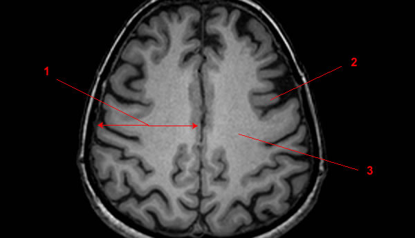
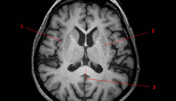
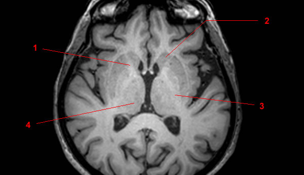
The distribution white matter – gray matter inside the brain is illustrated by MRI of the brain (axial sections). On some axial cuts, the thalamus and some basal ganglia (putamen, caudate nucleus, substantia nigra) were given as an example of “deep gray matter”. On other cuts, the corpus callosum and internal capsule have been indicated as an example of “white matter tracts”.

Several studies explored different approaches of using magnetic resonance imaging (MRI) in studying the white and gray matter of the brain(1).
These white and gray matters of the brain are located in the cerebral cortex of the brain’s cerebrum (the largest portion of the brain) that contains millions of glia and neurons(2).
The size, integrity, and shape of these white and gray matters of the brain are quantitatively and qualitatively described in a structural MRI(3). Additionally, the structural MRI shows the information on the anatomical visualization of the brain’s regions and patterns(4).
Generally, the brain’s white matter sends information through the nerve fibers (axons) in the brain, while the gray matter processes this information(5). Moreover, these tissues have other characteristics and roles in the functioning of the brain.
White Matter of the Brain
In the subcortical part (deeper tissues) of the brain lies the white matter that contains axons or nerve fibers enclosed with myelin(6).
The myelin sheath protects the nerve fibers from injury, enhances the transmission and communication of the electrical nerve signals in the axons, and gives the white matter color(7).
The white matter is responsible for coordinating communication, modulating action potentials, and relaying information between the brain’s different areas(8). It is primarily associated with the cognition and processing of information in the brain(9).
Corpus callosum is the most extensive white matter in the brain. It facilitates the interhemispheric communication between the left and right cerebral hemispheres(10).
There are three bundles of axons or tracts in the white matter that connect the different parts of the brain(11). These tracts are the commissural tract, association tract, and the projection tract(12).
The projection tract is vertically located between the lower and higher brain areas and the spinal cord centers(13). The tract is accountable for delivering information between the cerebrum and the rest of the body(14).
Crossing over from one cerebral hemisphere to the next through commissures or bridges is the commissural tract(15). This white matter tract allows the right and left sides of the cerebrum to communicate(16).
Finally, the association tracts link the brain’s various regions of the same hemisphere(17). Specifically, this tract connects the memory and perceptual centers of the brain(18).
Gray Matter of the Brain
The gray matter comprises most of the brain’s outer layer and has a high concentration of neuronal cell bodies (contains axons and dendrites)(19).
Neuron somas (the central part of the neuron where dendrites branch off) are primarily seen in the gray matter. When they are in circulation, neuron somas are tan in color. When examined outside the body, they are gray(20).
This gray matter runs from the brain to the spinal cord and forms a horn-like structure in the spinal column to enhance the gray matter’s signaling(21).
Processing and releasing information can be done due to the many neurons present in the gray matter(22). This process is possible through the communication of the axons in the white matter of the brain(23).
The gray matter also modulates memory, emotions, and movement of an individual(24). It has three sections: the anterior (anterior horn of the spinal cord), posterior (receives sensory information), and lateral gray column (receives input in the brain stems, hypothalamus, and organs)(25).
Significance of Magnetic Resonance Imaging (MRI) of the Brain
One imaging technique used in studying the brain is magnetic resonance imaging (MRI). This procedure employs radio waves and powerful magnets to produce three-dimensional anatomical images(26).
MRI is a non-invasive way of diagnosing and detecting diseases and monitoring treatment(27). According to an article published in the National Institutes of Health, MRI provides more explicit and detailed images of the brain, the spinal cord, nerves, tendons, and ligaments compared to computed tomography (CT) scans and X-rays(28).
A study suggested that tissue clustering in MRI analysis might pose possible advantages due to its partial volume effects, accountability, and simplicity(29).
Moreover, this MRI analysis method provides radiologists the flexibility to monitor and analyze specific brain regions(30).
A functional MRI (fMRI) can determine specific parts of the brain and the location where certain bodily functions, like memory, ensue(31). This imaging technique can also evaluate the effects of stroke and examine each part of the brain and its functional anatomy(32).
An fMRI procedure’s ability to determine the precise location of the brain’s functional center can aid physicians in planning treatments and surgeries for a specific brain condition or disorder(33).
- Harnsberger HR, Osborn AG, Ross JS, Moore KR, Salzman KL, Carrasco CR, Halmiton BE, Davidson HC, Wiggins RH. Diagnostic and Surgical Imaging Anatomy: Brain, Head and Neck, Spine. 3rd ed. Salt Lake City, Utah. Amirsys. 2007.
- Bourjat P, Veillon F. Imagerie radiologique tête et cou. Paris, Vigot. 1995.
- Gouazé A, Baumann JA, Dhem A. Sobota. Atlas d’Anatomie humaine. Tome 3. Système nerveux central, système nerveux autonome, organe des sens et peau, vaisseaux et nerfs périphériques. 1er éd. Paris, Maloine. 1977.
- Kahle W, Cabrol C. Anatomie. Tome 3: Système nerveux et organe des sens. 1er éd. Paris, Flammarion. 1979.
- Salih, Q. A., Ramli, A. R., Mahmud, R., & Wirza, R. (2005). Brain white and gray matter anatomy of MRI segmentation based on tissue evaluation. MedGenMed : Medscape general medicine, 7(2), 1.
- American Association of Neurological Surgeons, (n.d.), Anatomy of the Brain, retrieved from https://www.aans.org/en/Patients/Neurosurgical-Conditions-and-Treatments/Anatomy-of-the-Brain
- University of California San Diego School of Medicine, (n.d.), Structural MRI Imaging, retrieved from https://cfmriweb.ucsd.edu/Howto/3T/structure.html#:~:text=Broadly%20speaking%2C%20MRI%20signal%20varies,along%20with%20supporting%20glial%20cells.
- Ibid.
- Khan Academy, (n.d.), Gray and White Matter, retrieved from https://www.khanacademy.org/test-prep/mcat/behavior/biological-basis-of-behavior-ner/v/gray-and-white-matter#:~:text=In%20short%3A%20the%20axons%20(white,the%20conduction%20of%20neural%20activity.
- Medline Plus, (n.d.), White matter of the brain, retrieved from https://medlineplus.gov/ency/article/002344.htm
- Ibid.
- Lumen Boundless Anatomy and Physiology, (n.d.), The Cerebrum, retrieved from https://courses.lumenlearning.com/boundless-ap/chapter/the-cerebrum/#:~:text=White%20matter%20is%20composed%20largely,parts%20of%20the%20spinal%20cord.
- Ibid.
- Ibid.
- Ibid.
- Ibid,
- Ibid.
- Ibid.
- Ibid.
- Ibid.
- Ibid.
- Ibid.
- Mercadante AA, Tadi P. Neuroanatomy, Gray Matter. [Updated 2020 Jul 31]. In: StatPearls [Internet]. Treasure Island (FL): StatPearls Publishing; 2020 Jan-. Available from: https://www.ncbi.nlm.nih.gov/books/NBK553239/
- Ibid.
- Ibid.
- Ibid.
- Ibid.
- Ibid.
- Ibid.
- NIH National Institutes of Biomedical Imaging and Bioengineering, (n.d.), Magnetic Resonance Imaging, retrieved from https://www.nibib.nih.gov/science-education/science-topics/magnetic-resonance-imaging-mri
- Ibid.
- Ibid.
- Salih, Q. A., Op. Cit.
- Ibid.
- NIH National Institutes of Biomedical Imaging and Bioengineering, Op. Cit
- Radiology Info, (n.d.), Magnetic Resonance, Functional (fMRI) – Brain, retrieved from https://www.radiologyinfo.org/en/info.cfm?pg=fmribrain
- Ibid.