

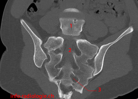
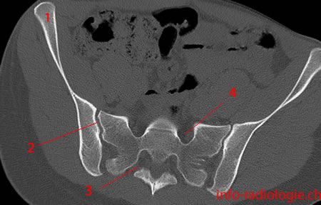
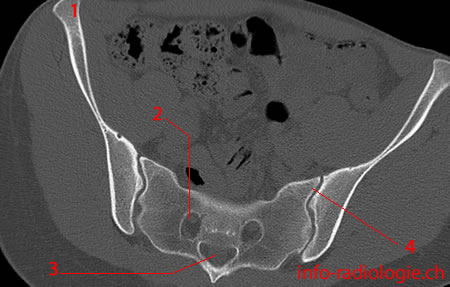
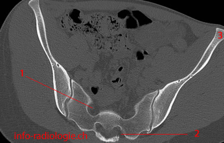
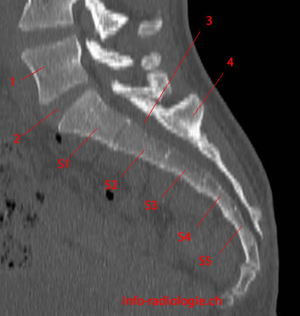
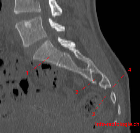
This photo gallery presents the anatomy of sacrum and coccyx by means of 3D-reconstructions, axial, sagittal and coronal reconstructions obtained from a scan of pelvis.
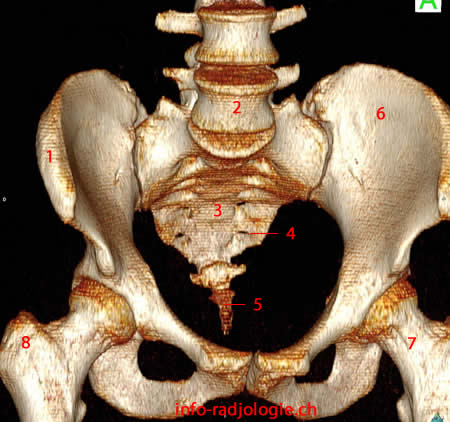
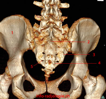

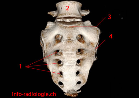

Sacrum
The sacrum refers to the bony structure located at the base of the lumbar vertebrae. It forms the posterior pelvic wall(1). The sacrum helps strengthen and stabilize the pelvis(2).
Women have a shorter sacrum than men(3). The female sacrum is distributed more obliquely backward, increasing the size of the pelvic cavity. Thus, the sacrum helps women endure pregnancy and offer more space for the developing fetus.
As it must be locked into the pelvis between the two ilia, the sacrum is massive(4). The bulk of the sacrum lies in the bodies and transverse elements of the upper two segments and the upper part of the third segment.
These segments enable the sacrum to be locked into the pelvic girdle and transfer axial forces laterally into the lower limbs and vice versa(5).
Fractures of the sacrum usually occur with high-energy trauma in young adults and low-energy trauma in the elderly or osteoporotic(6).
These fractures may be linked to other pelvic ring fractures. They may present with neurologic symptoms, such as bladder or bowel dysfunction and ipsilateral (the same side) leg weakness.
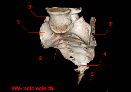
Coccyx
The coccyx refers to the terminal segment of the spine(7). It is a triangular bone composed of three to five fused segments. The largest segment articulates with the lowest sacral segment.
Although relatively small, the coccyx has several essential functions(8). It serves as the insertion site for various muscles, ligaments, and tendons.
Along with the ischial tuberosities, the coccyx provides weight-bearing support to individuals in the seated position(9). Leaning back while in a seated position causes increased pressure on the coccyx.
The coccyx provides positional support to the anus(10). However, the coccyx’s location makes it prone to internal injury during childbirth, particularly during a difficult or instrumented delivery.
Moreover, repetitive or prolonged sitting on hard or uncomfortable surfaces can cause minor trauma(11).
Coccydynia or tailbone pain refers to pain in and around the small triangular bone at the bottom of the spinal column (coccyx)(12). Tailbone pain may range from a dull ache to a fierce stab. The pain can last for weeks or months(13).
Various types of events can cause tailbone pain. External trauma can occur from a bruised or dislocated coccyx from a fall.
Meanwhile, internal trauma comes from difficult childbirth or from sitting on hard surfaces. Others include abscesses, infections, and tumors(14).
Women are five times more likely to obtain coccydynia than men(15). Moreover, adults and adolescents experience coccydynia more often than children.
Obese individuals are three times more prone to the condition(16). Those who lose weight fast may also be vulnerable to coccydynia.
Ways to Diagnose Sacrum and Coccyx Fractures
Radiographs
Plain anteroposterior and radiographs of the pelvis, sacrum, and lumbar spine are usually the first examinations to detect fractures involving the sacrum and coccyx(17). However, they can only detect complete fractures.
Early radiographs are often inconclusive because of fecal material, vascular calcifications, and bowel gas, which may overshadow the underlying fracture line(18).
In some instances, the fracture becomes evident only when the healing process is ongoing.
Magnetic Resonance Imaging
Magnetic resonance imaging (MRI) utilizes a magnetic field and computer-generated radio waves to generate detailed images of body organs and tissues(19).
MRI is one of the most sensitive screening techniques(20). It is also considered the standard diagnostic tool for sacral stress fractures.
A 2012 study published in the European Spine Journal examined the use of MRI in detecting painful adult coccyx(21). The researchers recommended the MRI of the painful coccyx when dynamic radiography failed to detect a pathological lesion.
Lesion refers to normal or slightly increased mobility of the coccyx or a rigid coccyx lacking a spicule.
Computed Tomography
Computed tomography (CT) refers to a diagnostic imaging test that generates detailed images of internal organs, bones, tissues, and blood vessels(22).
CT is both sensitive and specific(23). This procedure is a valid alternative to MRI in localizing the fracture line. It can produce accurate images that highlight sclerotic healing or fresh fracture lines.
Moreover, CT can be used together with MRI to rule out malignancy and osteomyelitis (infection of the bone).
How to Prevent Sacrum and Tailbone Injury
Consulting with healthcare professionals allows patients to avoid sacrum and tailbone problems. The following tips can also help them take care of the sacrum and coccyx.
- Maintain good posture. One should avoid slouching when sitting to avoid excessive pressure on the lumbosacral spine and the sacroiliac joints (joints connecting the sacrum to the left and right sides of the pelvis).
- Exercise regularly. Walking, jogging, swimming, and taking yoga classes can help keep the spine strong and healthy.
- Build core muscle strength. Excellent core muscle strength can help stabilize the sacrum.
- Always wear seatbelts. Car accidents may be a cause of lower spine trauma.