

The posterior cerebral arteries arise from the division of Basilar artery. The posterior cerebral arteries end above the tentorium, in the calcarine sulcus.
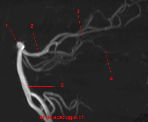
Posterior cerebral artery is divided into 4 parts:
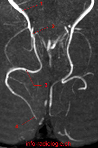
P1 segment courses from the basilar artery to the posterior communicating artery. The P1 segment passes over the oculomotor nerve (III).
P2 segment begins from the junction P1/posterior communicating artery. This segment curves around the midbrain (ambient cistern) to go near the trochlear nerve (IV), above the tentorium cerebelli.
P3 segment travels briefly through the quadrigeminal cistern
P4 segment ends in the calcarine fissure.
The posterior communicating arteries connect the arteries cérbrales posterior (P1/P2 junction) with the supraclinoid carotid arteries
The posterior cerebral arteries irrigate:
- midbrain
- thalamus
- posterior limb of internal capsule
- optic tract
- choroid plexus and cerebral peduncles
- splenium of corpus callosum
- 1/3 posterior medial of the cerebral hemispheres
- the region inferomedial temporal lobe and a large part of the occipital lobe.

- Dimmick SJ, Faulder KC. Normal variants of the cerebral circulation at multidetector CT angiography. Radiographics. 2009 Jul-Aug;29(4):1027-43.
- Harnsberger HR, Osborn AG, Ross JS, Moore KR, Salzman KL, Carrasco CR, Halmiton BE, Davidson HC, Wiggins RH. Diagnostic and Surgical Imaging Anatomy: Brain, Head and Neck, Spine. 3rd ed. Salt Lake City, Utah. Amirsys. 2007.
- Bourjat P, Veillon F. Imagerie radiologique tête et cou. Paris, Vigot. 1995.
- Gouazé A, Baumann JA, Dhem A. Sobota. Atlas d’Anatomie humaine. Tome 3. Système nerveux central, système nerveux autonome, organe des sens et peau, vaisseaux et nerfs périphériques. 1er éd. Paris, Maloine. 1977.
- Kahle W, Cabrol C. Anatomie. Tome 3: Système nerveux et organe des sens. 1er éd. Paris, Flammarion. 1979.