

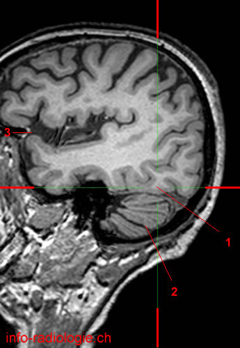
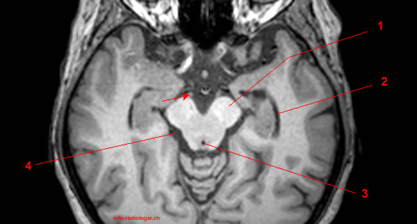
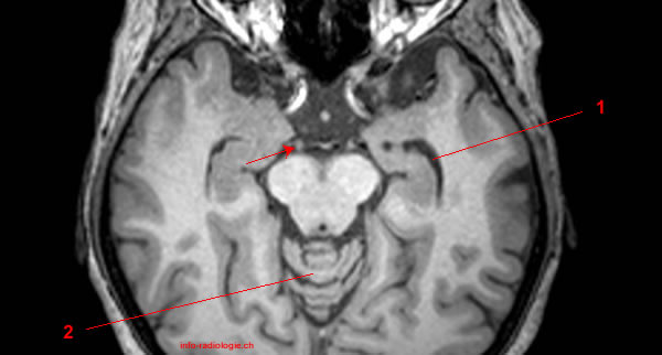
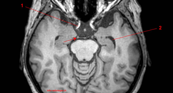
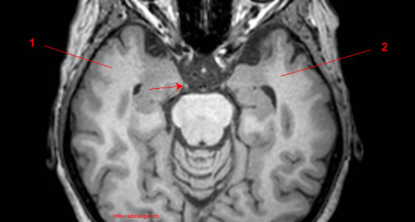
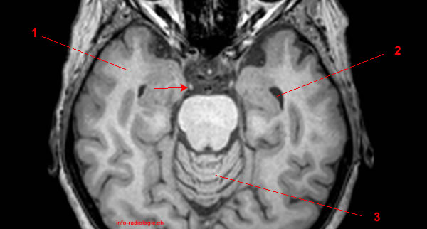
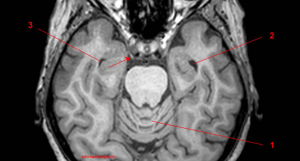
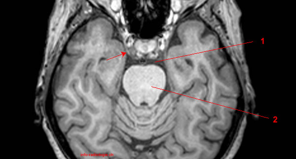
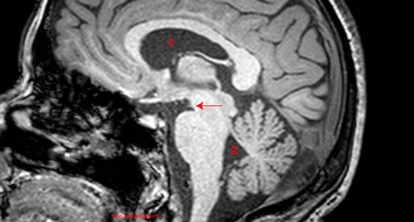
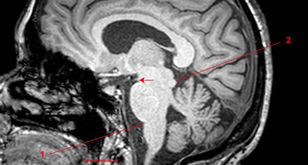
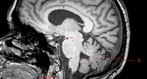
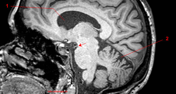
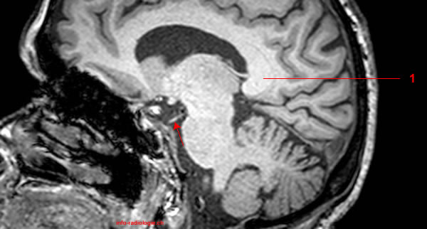
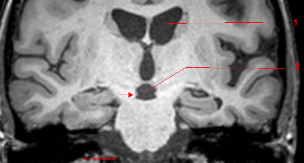
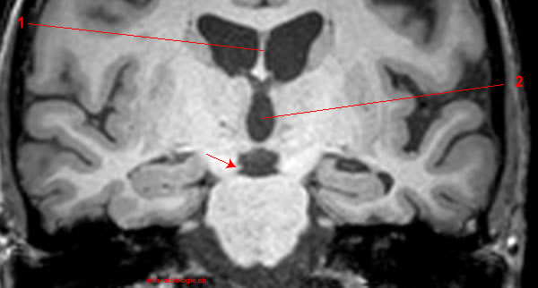
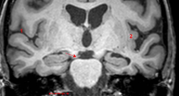
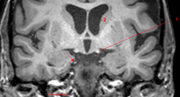
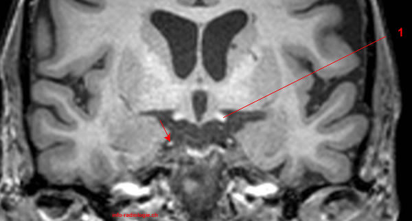
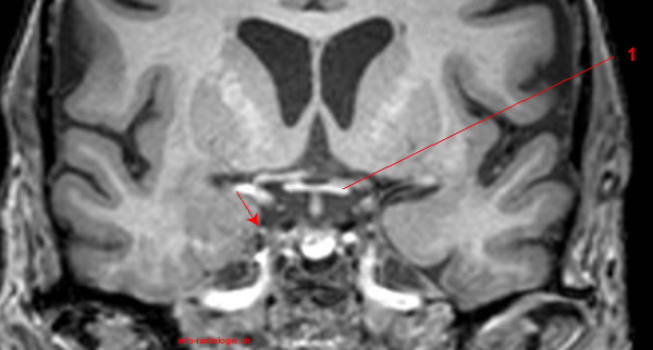
This page describes the path of the oculomotor nerve with brain MRI (axial, coronal and sagittal T1-weighted images).


Clinicians use magnetic resonance imaging (MRI) as the imaging technique to diagnose ischemic vasculopathy, mass formation, or aneurysm(1). MRI is also the modality of choice in patients with cranial nerve palsy and the assessment of ocular motor nerves(2-3).
The Oculomotor Nerve and Its Functions
The oculomotor nerve or the third cranial nerve is associated with eye motor functions(4). This nerve innervates to the upper eyelid (somatic), the pupil and lens (parasympathetic), and the eye muscles for gaze fixation and visual tracking (somatic)(5).
The oculomotor nerve originates from the oculomotor nucleus and accessory parasympathetic nucleus in the midbrain(6).
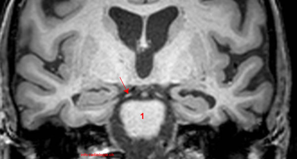
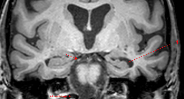
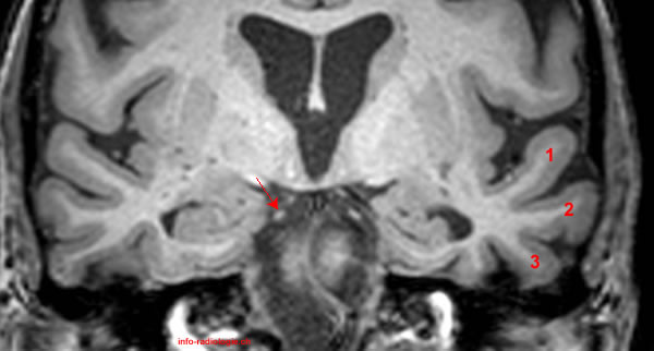
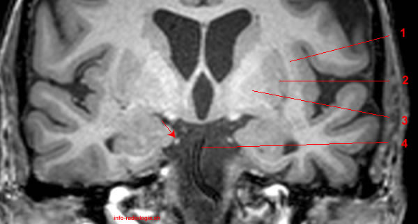
The oculomotor nerve exits the brainstem near the midline at the midbrain base and rear of the mammillary gland. The nerve passes through the supraorbital fissure (foramen or bony hollow in the skull) and cavernous sinus to reach the eye’s orbit(7).
There are two nerve fibers in the oculomotor nerve: the autonomic or involuntary fibers and the somatic or the voluntary fibers.
Somatic fibers are located deep inside the nerve, while the autonomic fibers are outside the nerve surrounding the somatic fibers(8).
Coordination of eye muscles for gaze fixation and visual tracking and the upper eyelid elevation are the oculomotor nerve’s voluntary functions (somatic)(9).
The oculomotor nerve innervates with four eye muscles to perform its voluntary functions. The inferior rectus muscle moves the eye down from the primary position, while the superior rectus muscle elevates the eye when looking straight ahead (primary position)(10).
The medial rectus muscle draws the eye towards the center from the primary position. The inferior oblique muscle elevates the eye when it is moved from the primary position(11).
The autonomic parasympathetic (involuntary) oculomotor nerve has innervations with sphincter pupillae (the smooth muscle near the pupils for pupil constrictions) and the ciliary muscles (the muscle connecting the iris to the choroid)(12).
Several movements assist the oculomotor nerves in coordination and adjustment of eye position(13).
The smooth pursuit helps the eye focus when there is a moving object, while the vestibulo-ocular reflexes adjust the eye movement during the head’s quick motions. The rapid and jerky eye motions are called saccades(14).
Furthermore, the optokinetic reflexes modulate the eye position to respond to a change in the visual field.
Oculomotor Nerve Dysfunctions
Due to its function in eye movement, the oculomotor nerve is a beneficial indicator of brain injury(15).
The most common causes of oculomotor nerve palsy or dysfunction are microvascular diseases, like hypertension, diabetes, tumor, trauma, or aneurysm(16).
Some of the clinical signs of oculomotor nerve palsy include diplopia or seeing double caused by the eye’s misalignment and droopy eyelids (ptosis).
Ptosis is caused by the muscle’s paralysis in the eye orbit that elevates the upper eyelid (levator palpebrae superioris)(17).
Oculomotor nerve compressions that involve superficial pupillomotor fibers and blood supply are due to lesions, such as tumors, aneurysms, and uncal herniation(18).
A scientific article cited that third-nerve palsy in children is acquired congenitally (43%), through trauma (20%), local inflammation (13%), and aneurysm (7%)(19).
Significance of MRI in the Assessment of Oculomotor Nerve
Magnetic resonance imaging is a non-invasive diagnostic procedure that produces a three-dimensional anatomical image. MRI is essential in treatment monitoring, disease detection, and diagnosis(20).
MRI uses strong magnetic fields and radio waves to evaluate muscles, the brain, tendons, ligaments, the spinal cord, and nerves more clearly(21).
MRI is the modality of choice when frequent imaging is required. This imaging technique does not use X-rays or radiation(22).
A specialized MRI called functional magnetic resonance imaging (fMRI) is often used to study the brain and observe its functions(23).
The fMRI gives an image of blood flow in the brain, brain activity, and brain structure’s functional anatomy(24). This procedure can detect brain abnormalities that cannot be found by other imaging techniques(25).
A study cited that an enhanced oculomotor nerve has been observed in an MRI test(26). The diagnostic imaging technique helped determine the connection between oculomotor nerve enhancement and vascular disease risk factors and inflammatory factors.
Moreover, a research study mentioned that MRI is the standard imaging technique for studying the cranial nerves and the diagnosis of nerve disorders(27).
A scientific article published in the American Journal of Roentgenology reviewed the accuracy of MRI in diagnosing peripheral nerve disease.
Results showed significant heterogeneity of MRI accuracy among studies. The most common MRI criteria in detecting abnormal peripheral nerves are nerve flattening, short tau inversion recovery (STIR) hyperintensity, and nerve enlargement(28).
Reference
• Harnsberger HR, Osborn AG, Ross JS, Moore KR, Salzman KL, Carrasco CR, Halmiton BE, Davidson HC, Wiggins RH. Diagnostic and Surgical Imaging Anatomy: Brain, Head and Neck, Spine. 3rd ed. Salt Lake City, Utah. Amirsys. 2007.
- Sethi, K. S., and Das, C. J., (June 2011), Magnetic Resonance Imaging of the Oculomotor Nerve, retrieved from https://journals.sagepub.com/doi/abs/10.1177/197140091102400316?journalCode=neub
- Ettl A, Salomonowitz E. Visualization of the oculomotor cranial nerves by magnetic resonance imaging. Strabismus. 2004 Jun;12(2):85-96. doi: 10.1080/09273970490517511. PMID: 15672931.
- Ferreira, T., Verbist, B., et. al., (May 2010), Imaging the Ocular Motor Nerves, retrieved from https://www.ejradiology.com/article/S0720-048X(10)00065-3/pdf
- Joyce C, Le PH, Peterson DC. Neuroanatomy, Cranial Nerve 3 (Oculomotor) [Updated 2020 Jul 27]. In: StatPearls [Internet]. Treasure Island (FL): StatPearls Publishing; 2020 Jan-. Available from: https://www.ncbi.nlm.nih.gov/books/NBK537126/
- Ibid.
- Ibid.
- Ibid.
- Ibid.
- Ibid.
- Ibid.
- Ibid.
- Ibid.
- Ibid.
- Ibid.
- Perry, C., (n.d.), Oculomotor Nerve, retrieved from https://www.kenhub.com/en/library/anatomy/the-oculomotor-nerve
- Joyce, C., Op. Cit.
- Ibid.
- Modi P, Arsiwalla T. Cranial Nerve III Palsy. [Updated 2020 Jul 10]. In: StatPearls [Internet]. Treasure Island (FL): StatPearls Publishing; 2020 Jan-. Available from: https://www.ncbi.nlm.nih.gov/books/NBK526112/
- Ibid.
- NIH National Institute of Biomedical Imaging and Bioengineering, (n.d.), Magnetic Resonance Imaging, retrieved from https://www.nibib.nih.gov/science-education/science-topics/magnetic-resonance-imaging-mri
- Ibid,
- Ibid.
- Ibid.
- Radiology Info, (n.d.), Magnetic Resonance, Functional (fMRI) – Brain, retrieved from https://www.radiologyinfo.org/en/info.cfm?pg=fmribrain
- Ibid.
- Yang, Y., Lai, C., Yan, F., & Wang, J. (2020). Clinical Significance of MRI Contrast Enhancement of the Oculomotor Nerve in Ischemic Isolated Oculomotor Nerve Palsy. Journal of clinical neurology (Seoul, Korea), 16(4), 653–658. https://doi.org/10.3988/jcn.2020.16.4.653
- Romano, N., Federici, M. & Castaldi, A. Imaging of cranial nerves: a pictorial overview. Insights Imaging 10, 33 (2019). https://doi.org/10.1186/s13244-019-0719-5
- Kwee, R. M., Chabbra, A., et al., (December 2014), Accuracy of MRI in Diagnosing Peripheral Nerve Disease: A Systematic Review of the Literature, retrieved from https://www.ajronline.org/doi/full/10.2214/AJR.13.12403