The Cervical Spine
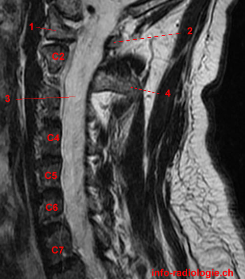
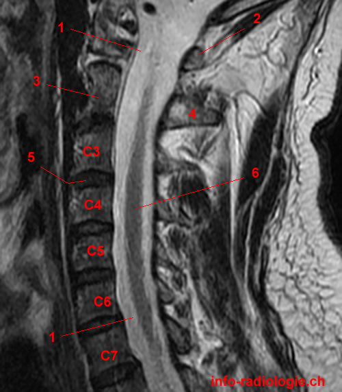
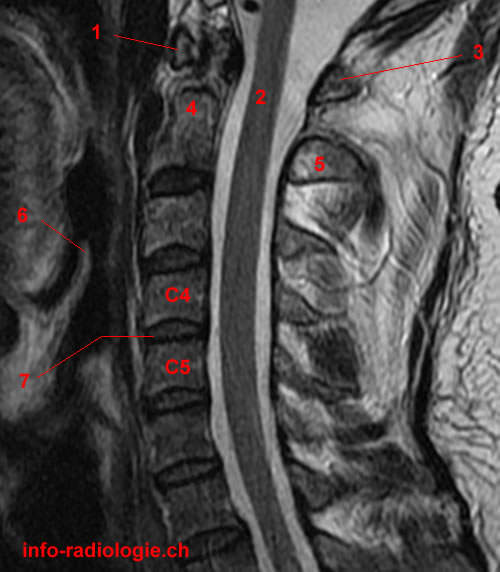
The neck is part of the spinal column or backbone, which extends through most of the body(1). The cervical spine or the neck region has seven bones separated from each other through intervertebral discs. These discs facilitate the free movement of the spine and serve as shock absorbers during activity.
The cervical spine allows the passage of essential vasculature (a network of blood vessels) to the brain and gives attachment sites for muscles that move the head, neck, and shoulder girdle(2).
Like the lumbar spine (lower back), the cervical spine has a lordotic curve or a backward C-shape(3). Compared with other spinal regions, the cervical spine is relatively more mobile.
There are special openings in each vertebra in the cervical spine for the arteries and the spinal canal that carries the spinal cord(4). The arteries, or blood vessels that take blood away from the heart, deliver blood through the brain as they run through the openings.
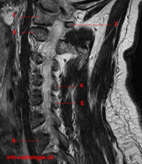
Magnetic Resonance Imaging of the Cervical Spine
The cervical spine needs to support the weight of the head. While the cervical spine is flexible, it has a high risk for injury from sudden, strong movements due to limited muscle support in the cervical area(5).
Magnetic resonance imaging (MRI) may be adopted to detect neck-related conditions. MRI of the cervical spine uses a magnetic field and radio waves to produce detailed images of the bones at the back of the neck(6).
Unlike computed tomography (CT) scan, MRI does not use radiation(7). Moreover, MRI produces better soft-tissue images than X-rays, allowing clinicians to accurately evaluate various body tissues, such as the spinal cord and vertebral disks. The procedure also helps identify healthy tissues from diseased ones.
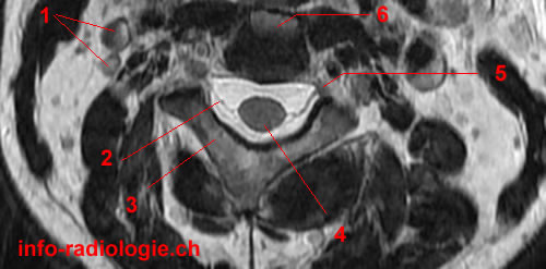
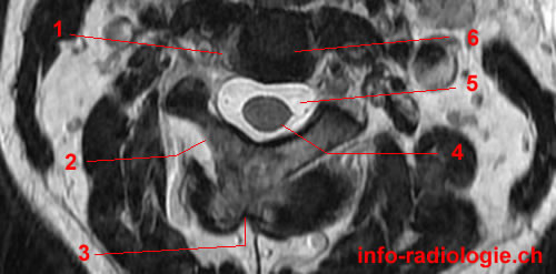
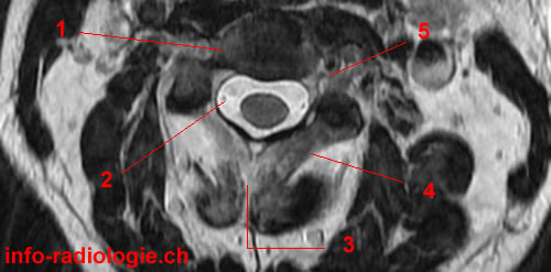
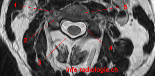
MRI may help detect different conditions involving the cervical spine and issues in the spinal column’s soft tissues, like the spinal cord, nerves, and disks(8).
The procedure is also used to assess injuries of the seven cervical spine bones(9). MRI may help evaluate pain, numbness, or weakness in the arms, shoulder, or neck area.
MRI can help detect chronic diseases of the nervous system(10). The test can diagnose tumors, swelling, bleeding, and inflammatory diseases in the vertebrate and surrounding tissues.
What to Expect From a Cervical Spine MRI
Before the Exam
An MRI of the cervical spine does not need special preparations(11). However, individuals taking the MRI exam should remove metal objects, like eyeglasses and jewelry, as they can produce a bright or blank spot on the diagnostic film. Braces and dental fillings do not interfere with the procedure.
The patients should inform the technologist, radiology nurse, or imaging physician of any allergies, ailments, previous drug interactions, or pregnancy.
Moreover, they need to tell the medical professionals if they have undergone back surgery or any other types of surgery.
Note that electronic devices are not allowed in the MRI room.
During the Exam
During the MRI exam, patients should stay still to get the highest quality MRI results. Given this, sedation may be necessary.
Sedation medications are often given through an intravenous (IV) placed on a vein. These medications may be beneficial for patients with claustrophobia (fear of enclosed areas)(12).
Moreover, a contrast solution may be given through an IV to highlight specific cervical spine problems, like infection or inflammation(13).
Cervical spine MRI often lasts for 30 to 45 minutes(14). Patients lie on the movable scanning table while a radiologic technologist places them into position.
A coil (a unique plastic device to enhance image quality) is placed above the patient’s neck(15). As the table slides into the tunnel, images of the neck are taken.
During the procedure, patients hear repetitive sounds from the machine, which is normal.
After the Exam
Patients may immediately resume their regular routines and diet, unless sedation is used or they are told to do otherwise(16).
MRI results are usually not given directly to the patient or their family at the time of the exam. The radiologist examines the MRI images and sends a report to the physician. The physician discusses the results of the procedure with the patient.
Risks of Cervical Spine MRI
As MRI does not use radiation, it does not pose any health risks to patients(17). Moreover, the procedure can be repeated without side effects.
However, sedation may pose some risks(18). Contrast solutions may also cause allergic reactions.
Nursing mothers should not breastfeed their babies 24 to 48 hours after contrast solutions are given(19). It is best to inform the physician of any allergies, drug interactions, or pregnancy to avoid possible complications.