

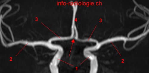
The Internal carotid artery divides into middle cerebral artery and anterior cerebral artery. The anterior cerebral artery enters the longitudinal interhemispheric fissure of the brain.
The anterior communicating artery connects right anterior communicating artery to left anterior communicating artery.
The anterior cerebral artery is divided into 3 parts:
A1 segment, horizontal, get around in the region of the optic nerve.
A2 segment, vertical, means the branch located in the interhemispheric fissure, up before the rostrum of the corpus callosum
A3 segment, distal, courses around the genu of corpus callosum. This segment A3 divides into pericallosal artery and callosomarginal artery.
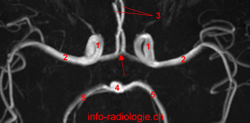
The anterior cerebral arteries irrigate:
- a large part of the medial region of the cerebral hemispheres (2/3 anterior)
- genu of corpus callosum
- anterior limb of the internal capsule
- head of nucleus caudatus.
What Is the Anterior Cerebral Artery?
Two vertebral arteries are responsible for the brain’s blood supply: the vertebrobasilar arteries or posterior circuit and the anterior circuit or the internal carotid arteries(1).
Vertebrobasilar arteries supply blood to the hindbrain and the occipital lobe (visual processing center of the brain). In contrast, the internal carotid arteries supply blood to the anterior (near the front) and the middle of the brain(2).
The anterior cerebral artery is the end branch or terminal branch of the internal carotid artery(3). A terminal branch or terminal artery lacks connection (anastomoses) with other arteries to avoid occlusion (blockage) of the blood supply and maintain blood circulation in the different tissues or organs(4).
The anterior cerebral artery connects with its opposite side (contralateral counterpart) through the anterior communicating artery, which is the short vessel that connects the left and right anterior cerebral artery(5).
This connection is an essential part of the cerebral circle or the circle of Willis(6). This circle is the anastomotic ring of arteries at the base of the brain(7).
From the medial end of the Sylvian fissure or lateral sulcus, the anterior cerebral artery extends rostrally (anterior) to the longitudinal fissure and continues around the genu of the corpus callosum(8).
The Sylvian fissure (lateral sulcus) separates the temporal lobes from the frontal and parietal lobes, while the longitudinal fissure is the deep groove that separates the two cerebral hemispheres. Meanwhile, the genu of the corpus callosum refers to the floor of the longitudinal fissure.
The anterior cerebral artery consists of two branches, namely the cortical and central branches(9). The central branches of the anterior cerebral artery supply blood to the deep cerebral structures(10).
Meanwhile, the cortical branch is further subdivided into three branches: the frontal branches, orbital branches, and the parietal branches(11).
The anterior cerebral artery’s course is divided into five segments: the precommunicating segment (A1), infracallosal segment (A2), precallosal segment (A3), supracallosal segment (A4), and the postcollosal segment (A5)(12).
Precommunicating Segment (A1)
This segment of the anterior cerebral artery is located at the internal carotid artery and runs to the anterior communicating artery(13). The segment lies inferiorly to the anterior perforated substance (the irregularly quadrilateral area in front of the optic tract) and superiorly situated to the optic chiasm (crossing of the optic nerves)(14).
The anterior communicating artery belongs to the precommunicating segment part of the anterior cerebral artery(15).
The anterior limb of the internal capsule and caudate nucleus are irrigated by the anterior communicating artery(16).
The caudate nucleus largely controls the voluntary skeletal movements, while the anterior limb of the internal capsule carries almost all the information that travels from and to the cerebral cortex.
Infracallosal Segment (A2)
The infracallosal segment is also called the vertical or postcommunicating segment(17). This segment traverses around the rostrum (part of the cranium that holds the nasal cavity, teeth, and palate) of the corpus callosum and spreads from the anterior of the communicating artery to the genu of the corpus callosum(18).
Precallosal Segment (A3)
This segment extends from the callosomarginal artery (the main branch of the distal part of the anterior cerebral artery), around the genu of the corpus callosum, and posteriorly goes above the rostral area of the corpus callosum’s body(19).
Supracallosal Segment (A4)
The supracallosal segment is one of the smaller branches of the anterior cerebral artery(20). This segment is located at the anterior corpus callosum and continues to the coronal suture, which separates the parietal bones from the frontal bone(21).
Postcollosal Segment (A5)
Another small branch of the anterior cerebral artery is the postcollosal segment, which passes the plane of the coronal suture and extends superiorly to the corpus callosum(22).
Angiography of the Anterior Cerebral Artery
The primary function of the anterior cerebral artery and its branches is to act as the main blood supply of the human frontal lobes(23).
Physicians and clinicians use angiography and other imaging techniques, such as computed tomography (CT) and magnetic resonance imaging (MRI), in studying arteries and blood vessels(24).
The computed tomography angiography (CTA) is used to assess and diagnose blood flow disorders and blood vessel-related diseases, like aneurysm, blockage, injuries, vessel ruptures or tears, tumors, and blood clots(25). This procedure is done through CT scanning of the tissues and blood vessels in the body(26).
Meanwhile, magnetic resonance angiography (MRA) does not use radiation like the CTA. Instead, MRA uses radio waves and magnetic fields to identify abnormalities and assess the blood vessels(27).
Unlike CTA, the MRA procedure does not require a contrast dye. Alternatively, MRA uses gadolinium-based contrast dye, which may pose a lesser risk for allergic reactions compared to the iodine-based contrast dye used in CTA(28).
- Dimmick SJ, Faulder KC. Normal variants of the cerebral circulation at multidetector CT angiography. Radiographics. 2009 Jul-Aug;29(4):1027-43.
- Harnsberger HR, Osborn AG, Ross JS, Moore KR, Salzman KL, Carrasco CR, Halmiton BE, Davidson HC, Wiggins RH. Diagnostic and Surgical Imaging Anatomy: Brain, Head and Neck, Spine. 3rd ed. Salt Lake City, Utah. Amirsys. 2007.
- Bourjat P, Veillon F. Imagerie radiologique tête et cou. Paris, Vigot. 1995.
- Gouazé A, Baumann JA, Dhem A. Sobota. Atlas d’Anatomie humaine. Tome 3. Système nerveux central, système nerveux autonome, organe des sens et peau, vaisseaux et nerfs périphériques. 1er éd. Paris, Maloine. 1977.
- Kahle W, Cabrol C. Anatomie. Tome 3: Système nnerveux et organe des sens. 1er éd. Paris, Flammarion. 1979.
- Crumbie, L., (November 2020), Arteries of the Brain, retrieved from https://www.kenhub.com/en/library/anatomy/arteries-of-the-brain
- Ibid.
- Gruijcic, R., (October 2019), Anterior Cerebral Artery, retrieved from https://www.kenhub.com/en/library/anatomy/anterior-cerebral-artery
- terminal artery. (n.d.) Farlex Partner Medical Dictionary. (2012). Retrieved January 20 2021 from https://medical-dictionary.thefreedictionary.com/terminal+artery
- Gruijcic, R., Op. Cit.
- Ibid.
- Ocran, E., (October 2020), Circle of Willis, retrieved from https://www.kenhub.com/en/library/anatomy/circle-of-willis
- Gruijcic, R., Op. Cit.
- Ibid.
- Ibid.
- Rea, P., (2015), Blood Supply of the Brain and Clinical Issues, retrieved from https://www.sciencedirect.com/science/article/pii/B9780128020302000066
- Gruijcic, R., Op. CIt
- IMAIOS Anatomical Parts, (n.d.), Precommunicating part; A1 segment – Pars precommunicalis; Segmentum A1, retrieved from https://www.imaios.com/en/e-Anatomy/Anatomical-Parts/Precommunicating-part-A1-segment
- Gruijcic, R., Op. Cit.
- IMAIOS Anatomical Parts, Op. Cit.
- Ibid.
- Gruijcic, R., Op. Cit.
- Ibid.
- Ibid.
- IMAIOS Anatomical Part, (n.d.), Anterior cerebral artery – Arteria cerebri anterior, retrieved from https://www.imaios.com/en/e-Anatomy/Anatomical-Parts/Anterior-cerebral-artery
- Gruijcic, R., Op. Cit.
- Ibid.
- Tahir, R. A., Haider, S., Kole, M., Griffith, B., & Marin, H. (2019). Anterior Cerebral Artery: Variant Anatomy and Pathology. Journal of vascular and interventional neurology, 10(3), 16–22.
- Makowicz, G., Poniatowska, R., & Lusawa, M. (2013). Variants of cerebral arteries – anterior circulation. Polish journal of radiology, 78(3), 42–47. https://doi.org/10.12659/PJR.889403
- Radiology Info, (n.d.), CT Angiography (CTA), retrieved from https://www.radiologyinfo.org/en/info.cfm?pg=angioct#:~:text=Doctors%20perform%20angiography%20using%3A,magnetic%20resonance%20imaging%20(MRI)
- Ibid.
- Radiology Info, (n.d), MR Angiography, retrieved from https://www.radiologyinfo.org/en/info.cfm?pg=angiomr
- Ibid.