

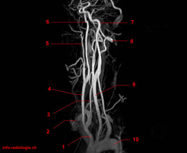
This gallery of images presents the anatomy of the neck vessels (carotid and vertebral arteries) by means of magnetic resonance angiography.
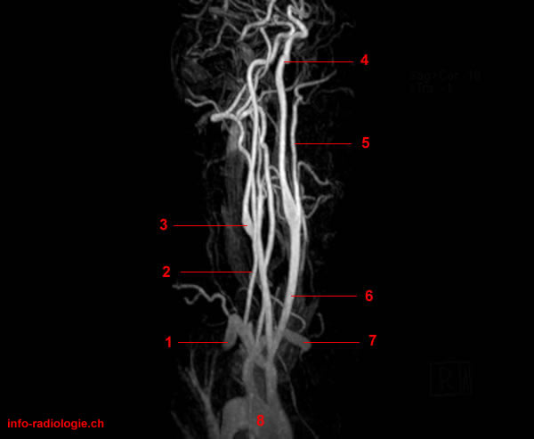
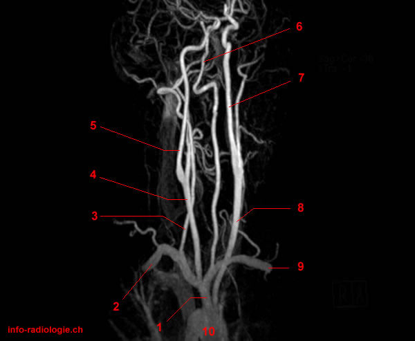
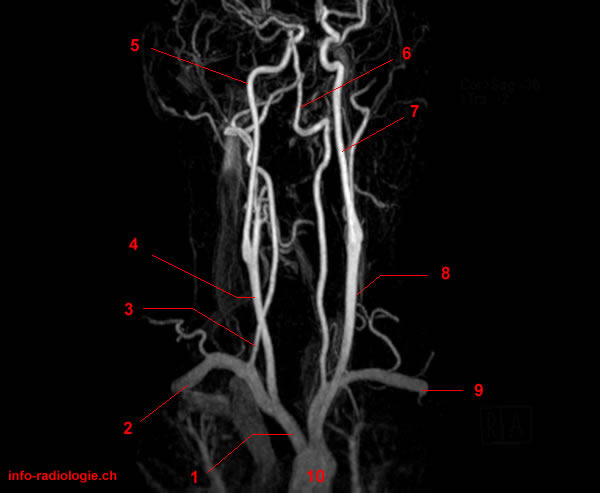
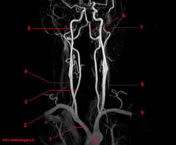
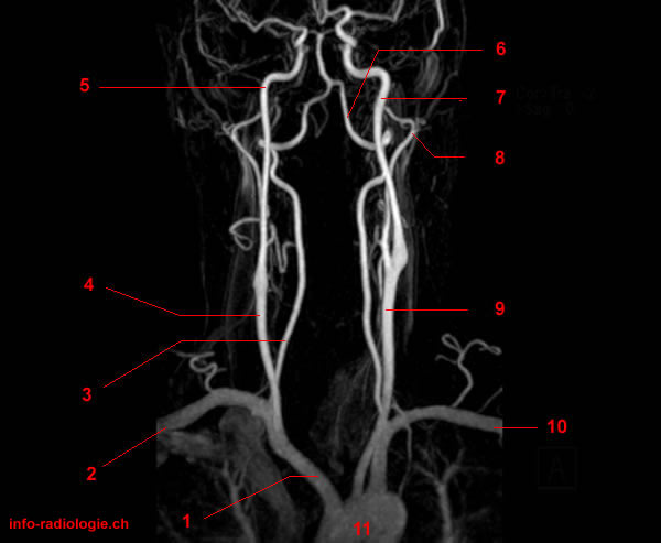
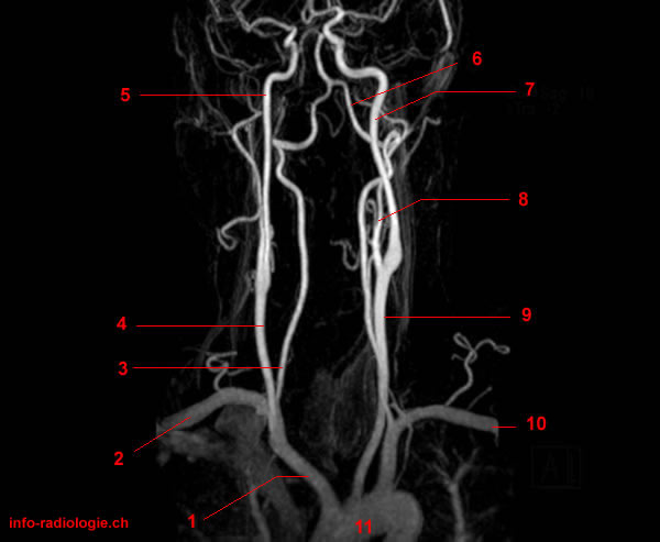
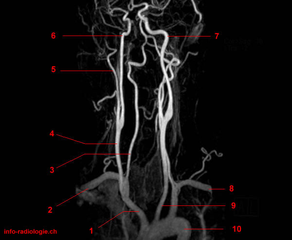
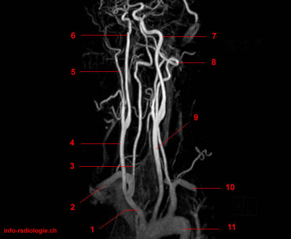
Carotid and Vertebral Arteries
Carotid and vertebral arteries lie on either side of the neck(1). They supply blood flow to the brain.
Carotid arteries consist of three layers: tunica intima (the inner layer), tunica media (the middle layer), and tunica adventitia (the outer layer(2). The tunica intima is composed of endothelium supported by a fragile elastic and a thick collagenous layer.
Meanwhile, the tunica media consists of smooth muscle cells(3). It helps change the diameter of the blood vessel to regulate blood flow and blood pressure.
The tunica adventitia attaches the carotid vessel to the surrounding tissue(4).
The carotid arteries originate posterior to the sternoclavicular joints (one of the joints that complete the shoulder)(5). In the neck, carotid arteries are contained within the carotid sheath posterior to the sternocleidomastoid muscle (one of the largest muscles).
Paired vertebral arteries bring blood supply to the upper part of the spinal cord, cerebellum, brainstem, and posterior part of the brain(6).
These arteries come from the first part of the subclavian artery. They move along the sides of the neck and merge at the pons to form the single midline basilar artery.
Occupying the back of the neck near the spine, the vertebral arteries cannot be felt during a physical exam(7).
MRA of Carotid and Vertebral Arteries
Dissection occurs when a tear in the artery wall causes blood to leak between the layers and separates them(8). Carotid or vertebral artery dissection refers to a dissection of the arteries on the neck. It may involve the carotid, vertebral, or multiple arteries.
Magnetic resonance angiography (MRA) and magnetic resonance imaging (MRI) may help diagnose the dissection(9).
MRA uses a magnetic field, radio waves, and a computer to examine blood vessels and identify abnormalities(10). This procedure does not use radiation and may require the injection of a contrast material, which helps improve images(11).
Compared with the contrast material used for computed tomography (CT), the one used for MRA is less likely to cause allergic reactions(12).
Meanwhile, MRI is an imaging modality that employs a magnetic field and computer-generated waves to form detailed images of the body’s organs and tissues(13).
A 2005 study published in the American Journal of Neuroradiology evaluated the sensitivity and specificity of contrast-enhanced MRA in detecting diseases in the carotid and vertebrobasilar systems(14).
Results showed 90% sensitivity and 97% specificity. The researchers noted that contrast-enhanced MRA accurately detects disease in the carotid vessels and vertebrobasilar circulation.
The authors added that the procedure may provide a comprehensive and noninvasive assessment of the head and neck arteries in a single study.
What to Expect From an MRA of the Carotid and Vertebral Arteries
Before the Examination
Patients undergoing the MRA exam should wear comfortable, loose-fitting clothing. They may have to change into a hospital gown for the exam.
Before the procedure, they should remove pieces of jewelry, piercing accessories, and other metal objects to ensure that these do not interfere with the scan.
Instructions about eating and drinking may vary per facility. However, unless the doctor provides special instructions, patients may follow their regular routines.
Patients should inform their doctors if they have allergies, illnesses, or other medications. Pregnant and nursing women should also inform their doctors beforehand to ensure that the baby is not harmed.
The doctor may suggest additional preparation based on the patient’s needs.
During the Examination
An MRA exam usually lasts for 30 to 90 minutes(15). Patients lie on the MRA table that moves through a hollow, donut-shaped machine.
A computer attached to the machine processes radio waves and magnetic fields to produce two-dimensional or three-dimensional images. The magnetic fields and radio waves are painless and harmless(16).
The technician may inject a contrast material into the patient’s hand or forearm to enhance the image’s quality(17).
Using speakers, the technician from another room can communicate with the patient.
The doctor may recommend an open MRA to claustrophobic patients and give them a calming medication.
After the Examination
A radiologist examines the images and sends a report to the doctor. The doctor shares the results with the patient.
A follow-up exam is necessary when a detected abnormality requires further assessment. It can also be performed to monitor if there have been changes in the abnormality.
Limitations of MRA
Compared with CT angiography, MRA cannot capture images of calcium deposits within the blood vessels(18). Moreover, MRA images of some arteries may not be as clear as catheter angiography images. In particular, MRA assessment of small vessels may be challenging.
Specific types of MRA machines have weight limits(19). Producing high-quality images depends on the patient’s ability to stay still and hold their breath when needed.
- Harnsberger HR, Osborn AG, Ross JS, Moore KR, Salzman KL, Carrasco CR, Halmiton BE, Davidson HC, Wiggins RH. Diagnostic and Surgical Imaging Anatomy: Brain, Head and Neck, Spine. 3rd ed. Salt Lake City, Utah. Amirsys. 2007.
- Bourjat P, Veillon F. Imagerie radiologique tête et cou. Paris, Vigot. 1995.
- Gouazé A, Baumann JA, Dhem A. Sobota. Atlas d’Anatomie humaine. Tome 3. Système nerveux central, système nerveux autonome, organe des sens et peau, vaisseaux et nerfs périphériques. 1er éd. Paris, Maloine. 1977.
- Kahle W, Cabrol C. Anatomie. Tome 3: Système nnerveux et organe des sens. 1er éd. Paris, Flammarion. 1979.
- Swedish. Carotid & Vertebral Ultrasound to Detect Atherosclerosis. Retrieved from https://www.swedish.org/services/neuroscience-institute/our-services/cerebrovascular-center/our-services/swedish-vascular-ultrasound/carotid-vertebral-duplex-exam#:~:text=The%20carotid%20and%20vertebral%20arteries,which%20develop%20inside%20artery%20walls
- Sethi D, Gofur EM, Munakomi S. Anatomy, Head and Neck, Carotid Arteries. [Updated 2020 Jul 27]. In: StatPearls [Internet]. Treasure Island (FL): StatPearls Publishing; 2020 Jan-. Available from: https://www.ncbi.nlm.nih.gov/books/NBK545238/
- Ibid.
- Ibid.
- Ibid.
- Vasković, J. Vertebral artery. Retrieved from https://www.kenhub.com/en/library/anatomy/vertebral-artery
- Cleveland Clinic. Cervical (Carotid or Vertebral) Artery Dissection. Retrieved from https://my.clevelandclinic.org/health/diseases/16857-cervical-carotid-or-vertebral-artery-dissection
- Ibid.
- Kasner, S. E., Hankins, L. L., Bratina, P., & Morgenstern, L. B. (1997). Magnetic resonance angiography demonstrates vascular healing of carotid and vertebral artery dissections. Stroke, 28(10), 1993–1997. doi.org/10.1161/01.str.28.10.1993. https://www.ahajournals.org/doi/full/10.1161/01.str.28.10.1993
- Radiology Info. MR Angiography (MRA). Retrieved from https://www.radiologyinfo.org/en/info.cfm?pg=angiomr
- Cedars Sinai. MRI & MRA Procedures. Retrieved from https://www.cedars-sinai.org/programs/heart/clinical/aortic/diagnostic-tests/mri-mra.html
- Radiology Info. Op Cit.
- Mayo Clinic. MRI. Retrieved from https://www.mayoclinic.org/tests-procedures/mri/about/pac-20384768
- Yang, C. W., Carr, J. C., Futterer, S. F., Morasch, M. D., Yang, B. P., Shors, S. M., & Finn, J. P. (2005). Contrast-enhanced MR angiography of the carotid and vertebrobasilar circulations. AJNR. American journal of neuroradiology, 26(8), 2095–2101. http://www.ajnr.org/content/26/8/2095
- Cedars Sinai. Op Cit.
- Ibid.
- Ibid.
- Radiology Info. Op Cit.
- Ibid.