Thigh Magnetic Resonance Imaging
The thigh has some of the body’s largest muscles. Thigh muscles are responsible for allowing normal gait and proper lower extremity function(1).
The medial thigh muscles are responsible for the adduction (movement of a body part toward the body’s midline) of the leg. Weak adductor muscles may cause knee instability and adductor strain(2).
Thigh muscles also protect neurovascular structures as they go through the proximal hip joint to the knee and lower leg(3).
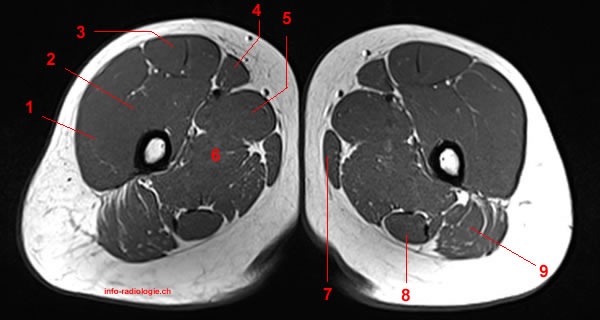
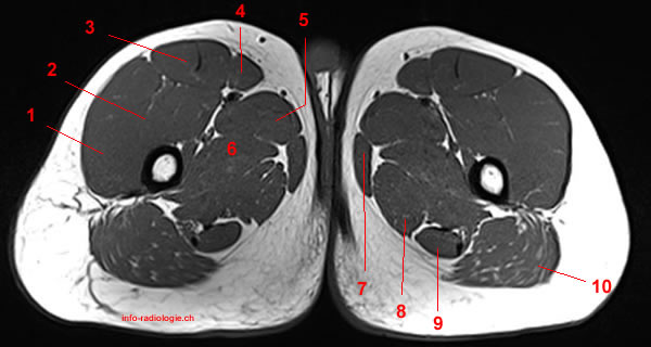
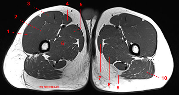
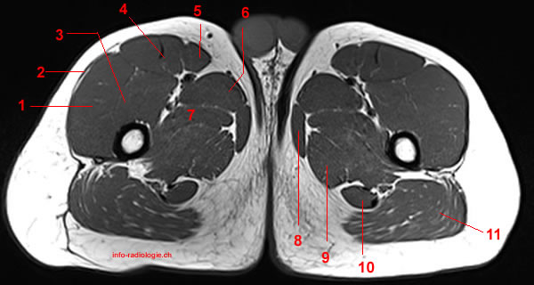
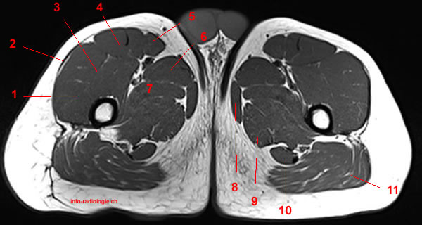
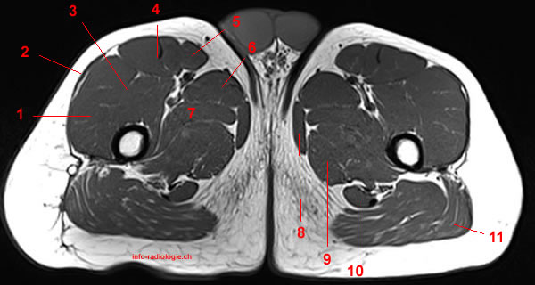
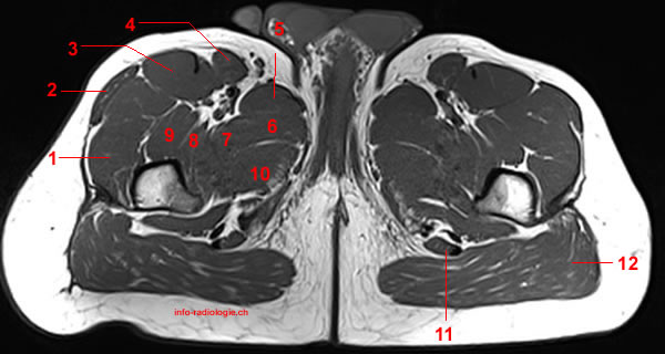
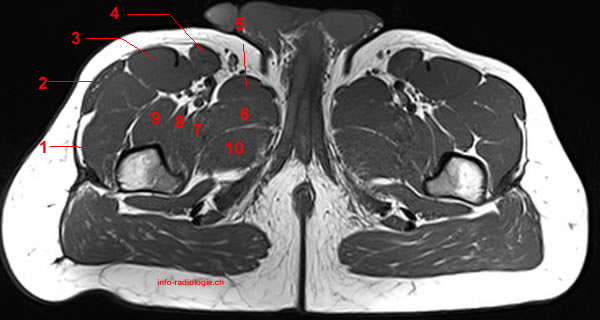
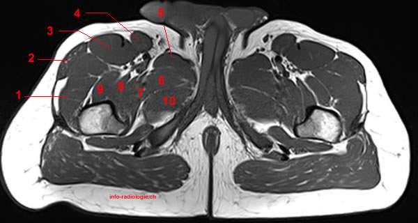
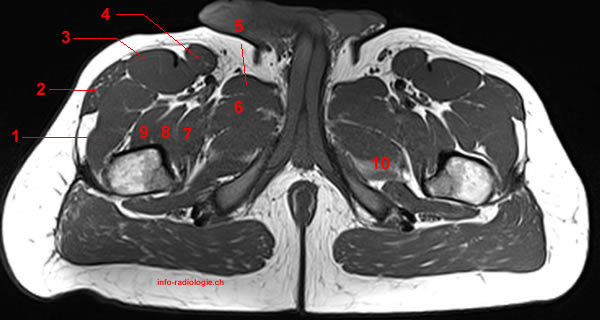
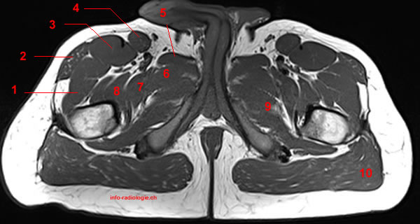
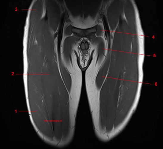
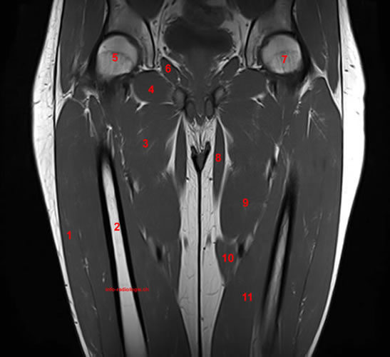
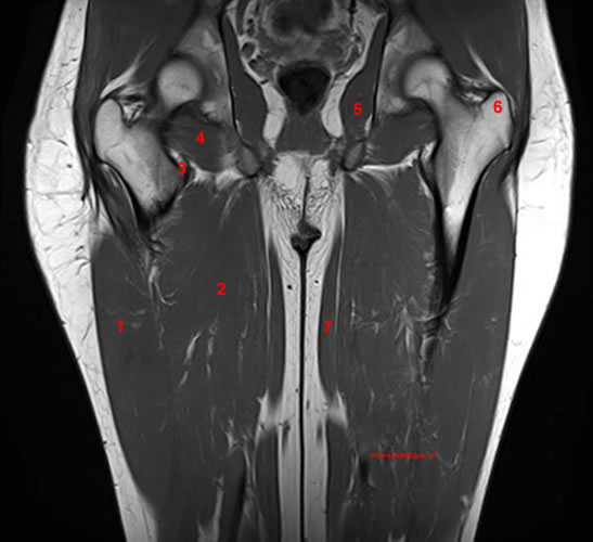
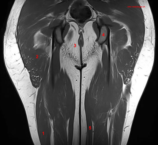
The thigh is composed of several muscles, including the quadriceps or quads (a group of four muscles)(4):
- Rectus femoris
- Vastus medialis
- Vastus lateralis
- Vastus intermedius
The rectus femoris is located in the center of the thigh, while the vastus medialis is in the middle of the said body part. Meanwhile, the vastus lateralis is on the side of the thigh, while the vastus intermedius is hidden below the rectus femoris(5).
The four muscles all extend the lower leg. The rectus femoris is also able to flex the thigh at the hip(6).
Magnetic resonance imaging (MRI) is a medical imaging procedure that may be used to diagnose conditions of the legs. It involves magnetic fields and radio waves to develop images of the body’s internal organs(7).
Other types of MRI, such as magnetic resonance angiogram (MRA), may capture medical images of the body’s blood vessels and blood flow(8).
Doing an MRA of the legs may help physicians detect stenosis (narrowing) and blockage of the arteries, also known as peripheral arterial disease. An MRA may also help surgeons prepare for surgery on the arteries of the legs(9).
What to Expect From a Thigh MRI
Before the Procedure
A patient undergoing thigh MRI may follow their routine and take medication as usual unless their physician advises otherwise.
Patients must inform their radiologist if they have any allergies to contrast dyes. They must also disclose if they are claustrophobic and in need of sedation.
Other important information to discuss with the doctor before the procedure includes the patient’s medical history and adverse reactions to prior medical imaging procedures. Patients must also inform their doctor if they are pregnant or wearing an on-body medication pump or pain patches.
Before undergoing an MRI, patients should inform their radiologist if they have a pacemaker, pain pump, defibrillator, loop recorder, stimulator, or other medical accessories. Informing the doctors about medical accessories helps avoid complications, as the MRI machine contains a powerful magnet.
During the Procedure
The patient may change into a hospital gown for the exam. They have to remove glasses, jewelry, and any metal objects in their possession that may interfere with the procedure.
If the MRI requires a contrast dye, the technologist may administer the dye through an intravenous administration. For non-contrast procedures, the patient skips this step.
The MRI technologist helps the patient lie on a table, which slides inside the center of a large scanner that has openings at both ends. The technician places an MRI coil over the patient’s thighs.
An MRI coil is a padded device that produces high-quality medical images of certain body parts(10).
In some instances, the MRI technician provides the patient with a squeeze ball. The squeeze ball serves as a way of getting in touch with the MRI technologist during the procedure.
If the patient has any questions or feels any discomfort during the MRI, they may squeeze the ball, and the attending technologist may help them with any issue they may be encountering.
The MRI machine makes a loud noise while sending out magnetic waves to create medical images. The table moves forward into the tube-like scanner and out when the scan has been completed.
A non-contrast MRI procedure should take 25 minutes. For an MRI exam with contrast media, the exam should take about 40 minutes to complete(11).
After the Procedure
Depending on the reason for the MRI test, the patient may be allowed to go home after the procedure. They can go back to their usual activities right away(12).
Risks of Thigh MRI
Exposure to strong magnetic fields during an MRI does not have any harmful effects on patients. However, the powerful magnet may interact with metal implants or other medical accessories inside the patient’s body(13).
There is a risk of an allergic reaction if contrast dye is used during the MRI procedure. However, most reactions are mild and treatable.
Contrast dyes for MRIs may contain the chemical gadolinium. Patients must inform their doctors of their allergies so they may avoid taking the contrast dye if they are allergic to gadolinium(14).
It is also recommended to inform the doctor if the patient is pregnant. Experts believe that very little contrast dye gets into breast milk, and even less may be passed on to the baby(15).
To avoid passing any contrast dye to their children, mothers should not breastfeed 24 hours after the procedure. They may use stored breast milk during the time they are flushing out the dye in their system.
15 Sources
References
- Ramage, J. & Varacallo, M. (2020). Anatomy, Bony Pelvis and Lower Limb, Medial Thigh Muscles. StatPearls. https://www.ncbi.nlm.nih.gov/books/NBK534775/
- Ibid
- Ibid
- Lumen Learning. Muscles of the hips and thighs. Retrieved from https://courses.lumenlearning.com/ap1x94x1/chapter/muscles-of-the-hips-and-thighs/
- Ibid
- Ibid
- North Oaks Health System. MRI of Upper Leg (Femur). Retrieved from https://www.northoaks.org/medical-services/diagnostics-imaging/mri/upper-leg/
- My Health Alberta. MRA of the Legs: About This Test. Retrieved from https://myhealth.alberta.ca/Health/aftercareinformation/pages/conditions.aspx?hwid=abk1324
- Ibid
- North Oaks Health System. Op cit.
- Ibid
- My Health Alberta. Op cit.
- Ibid
- Ibid
- Ibid